Abstract 抽象的
Ferroptosis is an iron-dependent cell death pathway that involves the depletion of intracellular glutathione (GSH) levels and iron-mediated lipid peroxidation. Ferroptosis is experimentally caused by the inhibition of the cystine/glutamate antiporter xCT, which depletes cells of GSH, or by inhibition of glutathione peroxidase 4 (GPx4), a key regulator of lipid peroxidation. The events that occur between GPx4 inhibition and the execution of ferroptotic cell death are currently a matter of active research. Previous work has shown that calcium release from the endoplasmic reticulum (ER) mediated by ryanodine receptor (RyR) channels contributes to ferroptosis-induced cell death in primary hippocampal neurons. Here, we used SH-SY5Y neuroblastoma cells, which do not express RyR channels, to test if calcium release mediated by the inositol 1,4,5-trisphosphate receptor (IP3R) channel plays a role in this process. We show that treatment with RAS Selective Lethal Compound 3 (RSL3), a GPx4 inhibitor, enhanced reactive oxygen species (ROS) generation, increased cytoplasmic and mitochondrial calcium levels, increased lipid peroxidation, and caused cell death. The RSL3-induced calcium signals were inhibited by Xestospongin B, a specific inhibitor of the ER-resident IP3R calcium channel, by decreasing IP3R levels with carbachol and by IP3R1 knockdown, which also prevented the changes in cell morphology toward roundness induced by RSL3. Intracellular calcium chelation by incubation with BAPTA-AM inhibited RSL3-induced calcium signals, which were not affected by extracellular calcium depletion. We propose that GPx4 inhibition activates IP3R-mediated calcium release in SH-SY5Y cells, leading to increased cytoplasmic and mitochondrial calcium levels, which, in turn, stimulate ROS production and induce lipid peroxidation and cell death in a noxious positive feedback cycle.
铁死亡是一种铁依赖性细胞死亡途径,涉及细胞内谷胱甘肽 (GSH) 水平的消耗和铁介导的脂质过氧化。铁死亡在实验上是由抑制胱氨酸/谷氨酸逆向转运蛋白 xCT(它会消耗细胞中的 GSH)或抑制谷胱甘肽过氧化物酶 4 (GPx4)(脂质过氧化的关键调节因子)引起的。 GPx4 抑制和铁死亡细胞死亡之间发生的事件目前是一个活跃的研究问题。先前的研究表明,兰尼碱受体(RyR)通道介导的内质网(ER)钙释放有助于原代海马神经元铁死亡诱导的细胞死亡。在这里,我们使用不表达 RyR 通道的 SH-SY5Y 神经母细胞瘤细胞来测试肌醇 1,4,5-三磷酸受体 (IP 3 R) 通道介导的钙释放是否在此过程中发挥作用。我们发现,用 RAS 选择性致死化合物 3 (RSL3)(一种 GPx4 抑制剂)治疗可增强活性氧 (ROS) 的产生,增加细胞质和线粒体钙水平,增加脂质过氧化,并导致细胞死亡。 RSL3 诱导的钙信号被 Xestospongin B(一种 ER 驻留 IP 3 R 钙通道的特异性抑制剂)抑制,通过用卡巴胆碱降低 IP 3 R 水平和通过 IP 3 R1 敲低,这也阻止了细胞形态向RSL3 诱导的圆度。通过与 BAPTA-AM 孵育的细胞内钙螯合抑制了 RSL3 诱导的钙信号,该信号不受细胞外钙消耗的影响。 我们认为,GPx4 抑制会激活 SH-SY5Y 细胞中 IP 3 R 介导的钙释放,导致细胞质和线粒体钙水平增加,进而刺激 ROS 产生,并在有害的正反馈循环中诱导脂质过氧化和细胞死亡。
Keywords: cell death, ferroptosis, calcium signaling, reactive oxygen species, endoplasmic reticulum, lipid peroxidation, oxidative stress, glutathione peroxidase, RSL3
关键词:细胞死亡, 铁死亡, 钙信号转导, 活性氧, 内质网, 脂质过氧化, 氧化应激, 谷胱甘肽过氧化物酶, RSL3
1. Introduction 一、简介
A major review, originating from the Nomenclature Committee on Cell Death, described 12 different types of cell death, which include intrinsic apoptosis, extrinsic apoptosis, membrane permeability transition (MPT)-dependent necrosis, necroptosis, ferroptosis, pyroptosis, parthanatos, entotic cell death, NETotic cell death, lysosome-dependent cell death, autophagy-dependent cell death, and immunogenic cell death [1]. Within these cell death processes, ferroptosis stands out, with over 4000 publications during 2023 alone.
来自细胞死亡命名委员会的一项重要评论描述了 12 种不同类型的细胞死亡,其中包括内在细胞凋亡、外在细胞凋亡、膜通透性转变 (MPT) 依赖性坏死、坏死性凋亡、铁死亡、细胞焦亡、parthanatos 和内溶性细胞死亡,NETotic细胞死亡,溶酶体依赖性细胞死亡,自噬依赖性细胞死亡和免疫原性细胞死亡[ 1 ]。在这些细胞死亡过程中,铁死亡最为突出,仅 2023 年就有 4000 多篇出版物发表。
Ferroptosis is a cell death process characterized by the accumulation of lipid peroxides in a manner that is dependent on cellular iron content [2,3]. Its importance, and the recent attention it has received, is related to the participation of ferroptosis in some of the most relevant of today pathologies, such as cancer and neurodegeneration [4,5]. Lately, the use of ferroptosis as a cell killing mechanism has brought about new strategies for the treatment of various malignant diseases [6].
铁死亡是一种细胞死亡过程,其特征是脂质过氧化物以依赖于细胞铁含量的方式积累[ 2 , 3 ]。它的重要性以及最近受到的关注与铁死亡在当今一些最相关的病理学中的参与有关,例如癌症和神经变性[ 4 , 5 ]。最近,利用铁死亡作为细胞杀伤机制为治疗各种恶性疾病带来了新的策略[ 6 ]。
The initial characterization of ferroptosis began in the context of a study using high-throughput screening to search for molecules with anti-cancer activity [7]; among other agents, erastin was found to produce non-apoptotic cell death much more efficiently in cells with the active RAS oncogene. Later insights into the effects of erastin determined its binding to mitochondrial voltage-gated anion channels (VDACs) [8] and excluded canonical cell death mechanisms, such as apoptosis, autophagic death, and necroptosis. Erastin-induced death is morphologically different from the classic types of cell death; it involves neither nuclear fragmentation nor vacuolization, and it entails a reduction in cytoplasm size, mitochondrial swelling, and plasma membrane rupture [8,9].
铁死亡的初步表征始于一项利用高通量筛选寻找具有抗癌活性的分子的研究[ 7 ];在其他药物中,erastin 被发现能够在具有活性 RAS 癌基因的细胞中更有效地产生非凋亡细胞死亡。后来对erastin作用的深入了解确定了它与线粒体电压门控阴离子通道(VDAC)的结合[ 8 ],并排除了典型的细胞死亡机制,例如细胞凋亡、自噬性死亡和坏死性凋亡。橡皮素诱导的死亡在形态上不同于典型的细胞死亡类型。它既不涉及核碎裂也不涉及空泡化,并且会导致细胞质尺寸减小、线粒体肿胀和质膜破裂[ 8 , 9 ]。
After these first studies, other compounds were found that induce the same type of cell death, with identical morphological characteristics and insensitivity to inhibitors, among others; the most notable among them was RSL3 [3]. Erastin and RSL3 induce a type of death with oxidative characteristics, which depends on iron, and it is characterized by the accumulation of lipid peroxides [9]. In particular, erastin, in addition to binding to VDAC channels, blocks the cystine/glutamate exchanger xCT, which leads to cellular GSH depletion [2,10]. This decrease in cellular GSH content leads to an oxidative imbalance that, among other factors, results in the inhibition of GPx4, which is the key enzyme that catalyzes the neutralization of membrane lipid peroxides to their respective alcohols. In contrast, RSL3 directly inhibits GPx4, resulting in the same accumulation of lipid peroxides but without decreasing GSH concentration [11,12,13,14].
在这些初步研究之后,发现其他化合物可诱导相同类型的细胞死亡,具有相同的形态特征和对抑制剂不敏感等;其中最引人注目的是 RSL3 [ 3 ]。 Erastin和RSL3诱导一种具有氧化特征的死亡,这种死亡依赖于铁,其特征是脂质过氧化物的积累[ 9 ]。特别是,erastin 除了与 VDAC 通道结合外,还能阻断胱氨酸/谷氨酸交换器 xCT,从而导致细胞 GSH 耗竭 [ 2 , 10 ]。细胞内谷胱甘肽含量的减少会导致氧化失衡,除其他因素外,还会导致 GPx4 受到抑制,而 GPx4 是催化膜脂过氧化物中和其各自醇的关键酶。相反, RSL3直接抑制 GPx4,导致相同的脂质过氧化物积累,但不会降低 GSH 浓度 [11,12,13,14 ] 。
Of note is the relationship between the inhibition of GPx4 and increased ROS production. A mediator between these two landmarks of ferroptosis is the pro-apoptotic protein Bid [15]; based on the observation that Bid inhibition preserves cell viability in RSL3-treated cells, the authors propose a sequence of events in which GPx4 inhibition enhances 12/15 LOX activity, which results in lipid peroxide formation and BID transactivation. In turn, activated Bid binds to mitochondria, decreasing their membrane potential and increasing ROS production [15]. Some research groups consider that ferroptosis is equivalent to oxytosis, a type of cell death described in hippocampal neurons [16], which was previously reported as a form of glutamate-mediated excitotoxicity [10]. In oxytosis, cell death may be initiated in a way similar to ferroptosis, that is, by blocking the xCT system. Since the xCT system is an exchanger-type co-transporter that uses the glutamate gradient to allow for cystine entry, when glutamate is present in excess (>5 mM) in the extracellular medium, it stops this gradient and inhibits cystine entrance. Since cystine is a GSH precursor, GSH concentrations decrease, which results in LOX activation, ROS generation, lipid peroxidation, and cell death. Death occurs in the absence of caspase activation, and there is protection by iron chelators. However, unlike what has been reported for ferroptosis, the execution of oxytosis depends on the massive influx of calcium from the extracellular medium [17]. The resulting intracellular calcium increase affects the mitochondria, producing the release of apoptosis-inducing factor (AIF), which can then translocate to the nucleus [18].
值得注意的是 GPx4 的抑制与 ROS 产生增加之间的关系。铁死亡这两个标志物之间的中介者是促凋亡蛋白 Bid [ 15 ];基于 Bid 抑制可保留 RSL3 处理细胞中细胞活力的观察结果,作者提出了一系列事件,其中 GPx4 抑制增强了 12/15 LOX 活性,从而导致脂质过氧化物形成和 BID 反式激活。反过来,激活的 Bid 与线粒体结合,降低其膜电位并增加 ROS 的产生 [ 15 ]。一些研究小组认为铁死亡相当于催眠,这是海马神经元中描述的一种细胞死亡类型[ 16 ],此前曾报道过这是谷氨酸介导的兴奋性毒性的一种形式[ 10 ]。在催眠过程中,细胞死亡可能以类似于铁死亡的方式启动,即通过阻断 xCT 系统。由于 xCT 系统是一种交换器型协同转运蛋白,它使用谷氨酸梯度来允许胱氨酸进入,因此当细胞外介质中谷氨酸过量(> 5 mM)时,它会停止该梯度并抑制胱氨酸进入。由于胱氨酸是 GSH 前体,GSH 浓度降低,导致 LOX 激活、ROS 生成、脂质过氧化和细胞死亡。死亡发生在没有半胱天冬酶激活的情况下,并且有铁螯合剂的保护。然而,与报道的铁死亡不同,催眠的执行取决于细胞外介质中钙的大量流入[ 17 ]。由此产生的细胞内钙增加会影响线粒体,产生凋亡诱导因子(AIF)的释放,然后该因子可以转移到细胞核中[ 18 ]。
The role of calcium in ferroptosis was dismissed by pioneering studies, due to the lack of protective effects of CoCl2 and GdCl3, which are both plasma membrane calcium channels blockers [19]. A report on HT-1080 fibrosarcoma cells [2] also dismissed the role of calcium in ferroptosis, arguing that neither the calcium indicator fura-2 nor intracellular calcium chelation with BAPTA-AM showed protection. In contrast, Maher’s group proposed the hypothesis that ferroptosis and oxytosis are the same phenomenon, reporting the protective effect of CoCl2 against RSL3- and erastin-induced cell death, in a manner analogous to that classically shown for oxytosis [20,21]. In the same line, protective effects of the calcium chelator BAPTA and of CoCl2 against erastin-induced cell death have been reported in HT22 and LUHMES [22].
由于 CoCl 2和 GdCl 3缺乏保护作用(质膜钙通道阻滞剂),钙在铁死亡中的作用被开创性研究驳回了[ 19 ]。一份关于 HT-1080 纤维肉瘤细胞的报告 [ 2 ] 也驳斥了钙在铁死亡中的作用,认为钙指示剂 fura-2 和细胞内钙与 BAPTA-AM 的螯合都没有显示出保护作用。相比之下,Maher 的小组提出了铁死亡和催产作用是相同现象的假设,报告了 CoCl 2对 RSL3 和erastin 诱导的细胞死亡的保护作用,其方式类似于经典的催产作用 [ 20 , 21 ]。在同一系列中,HT22 和 LUHMES 中报道了钙螯合剂 BAPTA 和 CoCl 2对erastin 诱导的细胞死亡的保护作用[ 22 ]。
Ferroptosis is accompanied by increased ROS levels; interestingly, many components that regulate intracellular calcium levels are stimulated by ROS, including the ER-resident calcium channels IP3R and RyR [23,24,25,26]. Thus, it is conceivable that the resulting calcium increase, originating from ROS-stimulated calcium release from the ER, could contribute to ferroptosis. In this line, a recent work reported that the suppression of calcium release mediated by RyR channels, through the use of inhibitory concentrations of ryanodine, offers significant but partial protection against RSL3-induced cell death in hippocampal neurons [27]. However, the putative role of IP3R-mediated calcium release in ferroptosis is unknown. Given this background, the role of calcium in ferroptosis is clearly a matter of debate and may have nuances that depend on the cell system studied.
铁死亡伴随着 ROS 水平升高;有趣的是,许多调节细胞内钙水平的成分都会受到ROS的刺激,包括内质网驻留钙通道IP 3 R和RyR [ 23,24,25,26 ]。因此,可以想象,由 ROS 刺激的内质网钙释放引起的钙增加可能会导致铁死亡。在这方面,最近的一项研究报告称,通过使用抑制浓度的兰尼碱抑制 RyR 通道介导的钙释放,可提供显着但部分的保护,防止海马神经元中 RSL3 诱导的细胞死亡 [ 27 ]。然而,IP 3 R 介导的钙释放在铁死亡中的假定作用尚不清楚。鉴于这种背景,钙在铁死亡中的作用显然是一个有争议的问题,并且可能存在细微差别,具体取决于所研究的细胞系统。
Here, we explored the hypothesis that in human neuroblastoma cells that do not express RyR channels, inhibition of the GPx4 enzyme activates IP3R-mediated calcium release, which results in increased cytoplasmic and mitochondrial calcium levels that contribute to the development of ferroptotic cell death.
在这里,我们探讨了这样的假设:在不表达 RyR 通道的人神经母细胞瘤细胞中,抑制 GPx4 酶会激活 IP 3 R 介导的钙释放,从而导致细胞质和线粒体钙水平增加,从而导致铁死亡细胞死亡。
2. Materials and Methods 2. 材料与方法
2.1. Cell Culture 2.1.细胞培养
SH-SY5Y cells (ATCC, CRL-2266) were cultured at 37 °C and 5% CO2 in DMEM/F12 media (Gibco, New York, NY, USA) supplemented with 10% FBS and 1% PenStrep (Gibco). For experiments, 80% confluence cultures were used.
SH-SY5Y 细胞(ATCC,CRL-2266)在补充有 10% FBS 和 1% PenStrep (Gibco) 的 DMEM/F12 培养基(Gibco,New York,NY,USA)中于 37 °C 和 5% CO 2下培养。实验中使用了 80% 汇合度的培养物。
2.2. Cell Viability 2.2.细胞活力
Viability was assessed with the Vybrant® MTT Cell Proliferation Assay Kit (V-13154; Thermo Fisher Scientific, Waltham, MA, USA) following the instructions of the manufacturer. Alternatively, cells were tested for propidium iodide (PI) permeability by incubation for 15 minutes (min) with 1 mg/mL of the fluorescence marker PI (Thermo Fisher Scientific) in modified Krebs buffer. Afterwards, PI red fluorescence was detected in random fields with a Nikon (Tokyo, Japan) TMS epifluorescence microscope.
按照制造商的说明,使用 Vybrant ® MTT 细胞增殖测定试剂盒(V-13154;Thermo Fisher Scientific,Waltham,MA,USA)评估活力。或者,通过在改良的 Krebs 缓冲液中与 1 mg/mL 荧光标记 PI (Thermo Fisher Scientific) 一起孵育 15 分钟 (min),测试细胞的碘化丙啶 (PI) 渗透性。随后,使用 Nikon(日本东京)TMS 落射荧光显微镜在随机视野中检测到 PI 红色荧光。
2.3. Cell Morphology 2.3.细胞形态学
After RSL3 treatment, cells were washed with modified Krebs buffer and imaged in a phase-contrast Nikon microscope. Subsequently, images were segmented using ImageJ version 1.54g (https://imagej.net/ij) creating binary masks. From the resulting segmentation images, cell area, perimeter, and circularity (a measure of roundedness) were determined.
RSL3 处理后,用改良的 Krebs 缓冲液洗涤细胞,并在相差尼康显微镜中成像。随后,使用 ImageJ 版本 1.54g ( https://imagej.net/ij ) 创建二进制掩模对图像进行分割。根据生成的分割图像,确定细胞面积、周长和圆度(圆度的度量)。
2.4. IP3R1 Downregulation and Mitochondrial Calcium Detection
2.4. IP 3 R1 下调和线粒体钙检测
Cover-glass-grown cells were transfected using Lipofectamine 2000 (Thermo Fisher Scientific) and DNA at a 1:3 ratio. Cells were incubated for 6 hours (h) with the Lipofectamine/DNA complex. For IP3R1 downregulation, cells were transfected with PTRIPZ (Dharmacon, Lafayette, CO, USA) carrying a shRNA sequence against the type-1 IP3R (IP3R1) isoform and a red fluorescent (RFP) reporter sequence. Forty-eight hours after transfection with PTRIPZ, cells were challenged with RSL3 (5 µM, 4 h), and intracellular calcium was determined as described below. After calcium recording, cells were imaged to individualize transfected cells and their morphology, including their roundness degree.
使用 Lipofectamine 2000 (Thermo Fisher Scientific) 和 DNA 以 1:3 的比例转染盖玻片生长的细胞。将细胞与 Lipofectamine/DNA 复合物一起孵育 6 小时 (h)。对于 IP 3 R1 下调,细胞用 PTRIPZ(Dharmacon,Lafayette,CO,USA)转染,该 PTRIPZ 携带针对 1 型 IP 3 R (IP 3 R1) 同工型的 shRNA 序列和红色荧光 (RFP) 报告序列。 PTRIPZ 转染后 48 小时,用 RSL3(5 µM,4 小时)攻击细胞,并按如下所述测定细胞内钙。钙记录后,对细胞进行成像以个体化转染细胞及其形态,包括其圆度。
For mitochondrial calcium sensing, cells were transfected with the plasmid pCMV CEPIA2mt (Addgene, https://www.addgene.org), carrying the mitochondrial calcium sensor Cepia2 [28]. Similarly, mitochondrial calcium levels in pCMV CEPIA2mt-transfected cells were determined 48 h after transfection.
对于线粒体钙传感,用携带线粒体钙传感器 Cepia2 的质粒 pCMV CEPIA2mt(Addgene,https: //www.addgene.org )转染细胞 [ 28 ]。类似地,转染后48小时测定pCMV CEPIA2mt转染细胞中的线粒体钙水平。
2.5. Western Blot Analysis
2.5.蛋白质印迹分析
After the corresponding treatments, SH-SY5Y cells were treated for 15 min with Radioimmunoprecipitation Assay (RIPA) lysis buffer supplemented with proteinase inhibitors (Waltham, MA, USA). After centrifugation, supernatants were collected, analyzed by SDS-polyacrylamide gel electrophoresis, and samples were wet transferred to nitrocellulose membranes. Membranes were blotted overnight at 4 °C using the following primary antibodies: rabbit anti-IP3R1 (1/1000, Cell Signaling Technology, Danvers, MA, USA), mouse anti-β-actin (1/5000, Sigma-Aldrich, St. Louis, MO, USA), rabbit anti-cleaved Caspase-3 (1/1000, Cell Signaling Technology), rabbit anti-Bcl-2 (1/1000, Santa Cruz Biotechnology, Dallas, TX, USA). For the detection of RyR channels, Western blot analysis of SH-SY5Y cells was performed as previously described [29]. Samples were separated by electrophoresis in 3.5–8% Tris-Acetate gels; gels were immersed in Tris-Tricine buffer and run for the first hour at 80 mV and then for the following 2 h at 100 mV. The protein bands were transferred to PDVF membranes (Millipore Corp. Burlington, MA, USA) and were incubated for 1 h at room temperature using as blocking solution Tris-buffered saline (TBS) with 5% fat-free milk for RyR2 and IP3R detection, or 5% bovine serum albumin (BSA) for RyR1 and RyR3 detection. The membrane was incubated overnight at 4 °C in blocking buffers with specific primary antibodies anti-RyR1 (1:500, generously provided by Dr. Vincenzo Sorrentino), anti-RyR2 (1:3000, MA3916, Invitrogen, Carlsbad, CA, USA), anti-RyR3 (1:2000, ab9082, Sigma Aldrich), and anti-IP3R (1:2500, PA1-901, ThermoFisher Scientific). Membranes were washed with saline and were then incubated for 2 h at room temperature with anti-rabbit or anti-mouse IgG antibodies coupled to radish peroxidase (1/5000, Thermo Fisher Scientific). Image acquisition was performed with the Image Lab software Version 6.0, Chemidoc TM MP System (Bio-Rad #12003154, Hercules, CA, USA).
相应处理后,SH-SY5Y 细胞用补充有蛋白酶抑制剂(Waltham,MA,USA)的放射免疫沉淀测定(RIPA)裂解缓冲液处理 15 分钟。离心后,收集上清液,通过SDS-聚丙烯酰胺凝胶电泳进行分析,并将样品湿转移至硝酸纤维素膜上。使用以下一抗在 4 °C 下对膜进行印迹过夜:兔抗 IP 3 R1(1/1000,Cell Signaling Technology,Danvers,MA,USA)、小鼠抗 β-肌动蛋白(1/5000,Sigma-Aldrich) ,圣路易斯,密苏里州,美国),兔抗 cleaved Caspase-3(1/1000,Cell Signaling Technology),兔抗 Bcl-2 (1/1000,圣克鲁斯生物技术公司,达拉斯,德克萨斯州,美国)。为了检测 RyR 通道,如前所述对 SH-SY5Y 细胞进行蛋白质印迹分析 [ 29 ]。样品在 3.5-8% Tris-Acetate 凝胶中通过电泳分离;将凝胶浸入 Tris-Tricine 缓冲液中,并在 80 mV 下运行第一小时,然后在 100 mV 下运行接下来的 2 小时。将蛋白质条带转移至 PDVF 膜(Millipore Corp. Burlington,MA,USA),并使用含有 5% 脱脂牛奶的 Tris 缓冲盐水 (TBS) 作为封闭液在室温下孵育 1 小时,用于 RyR2 和 IP 3 R检测,或5%牛血清白蛋白(BSA)用于RyR1和RyR3检测。将膜在带有特异性一抗抗 RyR1(1:500,由 Vincenzo Sorrentino 博士慷慨提供)、抗 RyR2(1:3000,MA3916,Invitrogen,卡尔斯巴德,加利福尼亚州,美国)的封闭缓冲液中于 4 °C 孵育过夜)、抗 RyR3 (1:2000、ab9082、Sigma Aldrich) 和抗 IP 3 R (1:2500,PA1-901,赛默飞世尔科技)。 用盐水洗涤膜,然后与与萝卜过氧化物酶偶联的抗兔或抗小鼠 IgG 抗体(1/5000,Thermo Fisher Scientific)在室温下孵育 2 小时。使用 Image Lab 软件 6.0 版 Chemidoc TM MP 系统(Bio-Rad #12003154,Hercules,CA,USA)进行图像采集。
2.6. ROS Detection 2.6。活性氧检测
The production of ROS was detected by incubation of cells with 5 µM H2DCFDA (Thermo Fisher Scientific), which emits fluorescence when oxidized to dichlorofluorescein (DCF) [30]. An often-unrecognized fact is that DHDCF is a highly selective hydroxyl radical sensor [31].
通过将细胞与 5 µM H2DCFDA (Thermo Fisher Scientific) 一起孵育来检测 ROS 的产生,该细胞在氧化为二氯荧光素 (DCF) 时会发出荧光 [ 30 ]。一个经常被忽视的事实是 DHDCF 是一种高度选择性的羟基自由基传感器 [ 31 ]。
2.7. Calcium Change Recordings
2.7.钙变化记录
For fura-2-based cytoplasmic calcium determinations, cells were loaded for 30 min with 3 µM fura-2-AM in modified Krebs buffer at 37 °C. Afterwards, cells were washed twice with PBS and were kept in dark for 10 min before starting the recordings. The emitted fluorescence at 510 nm was recorded in a fluorimeter, alternating the excitation between 340 and 380 nm. Recordings lasted 6 min, where the first 2 min corresponded to basal fluorescence determination. Then, 0.01% Triton X-100 was added in order to permeabilize the cell membrane and saturate the probe, and the record was continued for additional 2 min. Finally, 50 mM EGTA was added to determine the fluorescence of the calcium-unbound probe. Calcium concentrations were calculated using the Grynkiewicz equation [32]:
对于基于 fura-2 的细胞质钙测定,在 37 °C 的改良 Krebs 缓冲液中加入 3 µM fura-2-AM,将细胞上样 30 分钟。然后,用 PBS 洗涤细胞两次,并在开始记录之前在黑暗中保存 10 分钟。在荧光计中记录 510 nm 发射的荧光,在 340 和 380 nm 之间交替激发。记录持续 6 分钟,其中前 2 分钟对应于基础荧光测定。然后,添加0.01% Triton X-100以透化细胞膜并使探针饱和,并继续记录另外2分钟。最后,添加 50 mM EGTA 以确定未结合钙的探针的荧光。使用 Grynkiewicz 方程计算钙浓度 [ 32 ]:
| [Ca2+]i (nM) = Kd × [(R − Rmin)/Rmax − R)] × Sfb [Ca 2+ ] i (nM) = Kd × [(R − R min )/R max − R)] × Sfb |
where Kd = 145 nM is for fura-2 at 20 °C [33]; R is the ratio between the fluorescence produced by the excitation at 340 nm (bound) and 380 nm (unbound); Rmax corresponds to the fluorescence ratio emitted by the calcium-saturated probe (after Triton X-100 treatment) and Rmin is the ratio of fluorescence emitted by the calcium-depleted probe (EGTA); Sfb is the ratio of the fluorescence recorded at 380 (unbound probe) after Triton-X-100 and EGTA additions [34].
其中 Kd = 145 nM 是 fura-2 在 20 °C 时的值 [ 33 ]; R是340 nm(结合)和380 nm(未结合)激发产生的荧光之间的比率; R max对应于钙饱和探针(Triton X-100处理后)发射的荧光比率,R min对应于钙贫探针(EGTA)发射的荧光比率; Sfb 是添加 Triton-X-100 和 EGTA 后在 380(未结合探针)处记录的荧光的比率 [ 34 ]。
For fluo-3 based calcium imaging, cells were loaded for 30 min with 5 µM fluo-3-AM in modified Krebs buffer at 37 °C. Then, cells were washed twice with PBS and left to rest for 10 min before recordings. Recordings were performed in an LSM 710 Zeiss (Carl Zeiss AG, Oberkochen, Germany) confocal microscope using the 488 nm laser for fluo-3 excitation. Setup configuration was kept constant for every condition in each experiment.
对于基于 Fluo-3 的钙成像,细胞在 37 °C 下在改良的 Krebs 缓冲液中加入 5 µM Fluo-3-AM 上样 30 分钟。然后,用 PBS 洗涤细胞两次,并在记录前静置 10 分钟。使用 LSM 710 Zeiss(Carl Zeiss AG,Oberkochen,德国)共焦显微镜进行记录,使用 488 nm 激光进行 Fluo-3 激发。每个实验中的每个条件的设置配置都保持不变。
2.8. Lipid Peroxidation 2.8.脂质过氧化
To assess lipid peroxidation after RSL3 treatment, cells were loaded with 5 µM BODIPY C11 (Thermo Fisher Scientific) at 37 °C for 30 min. Then, cells were washed twice with PBS and fixed for 15 min with 4% paraformaldehyde at 4 °C. Subsequently, cells were washed three times with PBS and were mounted in microscope slides to be imaged in a LSM 710 Zeiss confocal microscope.
为了评估 RSL3 处理后的脂质过氧化情况,在 37°C 下向细胞上样 5 µM BODIPY C11 (Thermo Fisher Scientific) 30 分钟。然后,用 PBS 洗涤细胞两次,并在 4°C 下用 4% 多聚甲醛固定 15 分钟。随后,用 PBS 洗涤细胞 3 次,并安装在显微镜载玻片上,以便在 LSM 710 Zeiss 共聚焦显微镜中成像。
For detection of 4-hydroxy-2-nonenal (HNE)-protein adducts, cells were fixed with 4% paraformaldehyde (PFA), followed by a permeabilization with 0.1% Triton-X-100 for 30 min and blocked with 4% BSA for 30 min. Cells were incubated with anti-HNE (1/400, Abcam, Waltham, MA, USA) at 4 °C overnight, washed three times with PBS, and incubated with an Alexa 488 anti-mouse secondary antibody (1/400, Thermo Fisher Scientific). Cells were washed two times with PBS and mounted in microscope slides to be imaged in an LSM 710 Zeiss confocal microscope.
为了检测 4-羟基-2-壬烯醛 (HNE)-蛋白加合物,用 4% 多聚甲醛 (PFA) 固定细胞,然后用 0.1% Triton-X-100 透化 30 分钟,并用 4% BSA 封闭 30 分钟。 30分钟将细胞与抗 HNE (1/400, Abcam, Waltham, MA, USA) 在 4 °C 下孵育过夜,用 PBS 洗涤 3 次,并与 Alexa 488 抗小鼠二抗 (1/400, Thermo Fisher科学)。用 PBS 洗涤细胞两次,并安装在显微镜载玻片上,以便在 LSM 710 Zeiss 共聚焦显微镜中成像。
2.9. Statistical Analysis
2.9.统计分析
The Shapiro–Wilk test was used to determine the normal distribution of replicates. For the comparison of multiple experimental conditions, one-way ANOVA was used to test for differences in mean values, and Dunnett’s post hoc test was used for comparisons between mean values. For the comparison of two experimental conditions, the unpaired two-tail Student’s t test was used to compare differences between mean values. A p value < 0.05 was taken as statistically significant.
夏皮罗-威尔克检验用于确定重复的正态分布。对于多个实验条件的比较,使用单因素方差分析来检验平均值的差异,并使用 Dunnett 事后检验来检验平均值之间的比较。为了比较两个实验条件,使用不配对的双尾学生 t 检验来比较平均值之间的差异。 p值<0.05被视为具有统计显着性。
3. Results 3. 结果
3.1. RSL3 Induces Ferroptosis in SH-SY5Y Neuroblastoma Cells
3.1. RSL3 诱导 SH-SY5Y 神经母细胞瘤细胞铁死亡
The effects of RSL3 in inducing ferroptosis in SH-SY5Y neuroblastoma cells have not been systematically characterized to date [35], so we first determined the ferroptotic characteristics of RSL3-induced SH-SY5Y cell death.
迄今为止,RSL3 在诱导 SH-SY5Y 神经母细胞瘤细胞铁死亡中的作用尚未得到系统表征[ 35 ],因此我们首先确定了 RSL3 诱导 SH-SY5Y 细胞死亡的铁死亡特征。
3.1.1. RSL3 Treatment Induces Changes in Cell Morphology and Time- and Concentration-Dependent Cell Death
3.1.1. RSL3 治疗诱导细胞形态变化以及时间和浓度依赖性细胞死亡
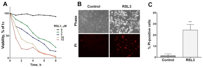
The viability of SH-SY5Y cells was reduced as a function of both the concentration and time of incubation with RSL3 (Figure 1A). In addition, RSL3 markedly affected cell morphology (Figure 1B, upper panel). Following RSL3 treatment, a number of cells detached from the substrate, showing a birefringent aspect (Figure 1B, top panels). The assessment of cell viability by PI stain showed a large increase in PI-stained non-viable cells in RSL3-treated cultures (Figure 1B, lower panels, and Figure 1C).
SH-SY5Y 细胞的活力随 RSL3 的浓度和孵育时间而降低(图 1 A)。此外,RSL3 显着影响细胞形态(图 1 B,上图)。 RSL3 处理后,许多细胞从基底上脱落,显示出双折射外观(图 1 B,顶图)。通过 PI 染色对细胞活力的评估显示,经 RSL3 处理的培养物中 PI 染色的非存活细胞大幅增加(图 1 B,下图和图 1 C)。
Figure 1. 图 1.
RSL3 affects the viability of SH-SY5Y cells. (A) Cells were treated with different concentrations of RSL3 for different times and viability was evaluated with the MTT assay. Viability at time = 0 was normalized to 100%. Values represent means of 5 replicates per experimental point of a representative experiment. N = 3 independent experiments. (B) Cells were treated for 3 h with 5 µM RSL3 or vehicle (DMSO, Control) and were stained with propidium iodine (PI, red) to evaluate dead cells in the population. Representative images are shown. Scale bar: 80 µm. (C) Quantification of PI-positive cells. Values represent the mean ± SD from 200–250 cells per experimental condition; *** p < 0.001.
RSL3 影响 SH-SY5Y 细胞的活力。 ( A ) 用不同浓度的 RSL3 处理细胞不同时间,并通过 MTT 测定评估活力。将时间 = 0 时的活力标准化为 100%。值代表代表性实验的每个实验点 5 次重复的平均值。 N = 3 个独立实验。 ( B ) 用 5 µM RSL3 或载体(DMSO,对照)处理细胞 3 小时,并用碘化丙啶(PI,红色)染色以评估群体中的死细胞。显示了代表性图像。比例尺:80 µm。 ( C ) PI 阳性细胞的定量。数值代表每个实验条件下 200-250 个细胞的平均值±SD; *** p < 0.001。
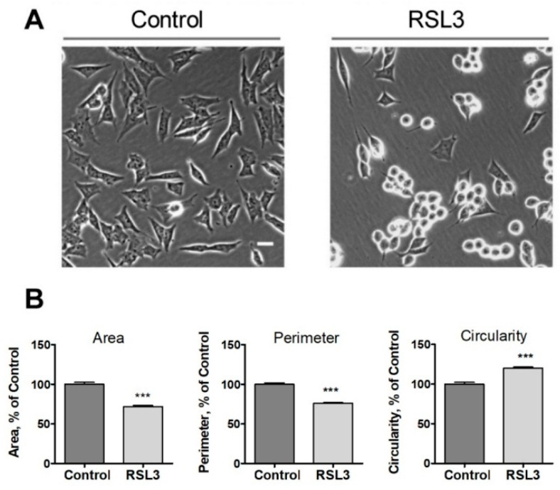
Following the above observations, we evaluated three morphological criteria that turned out to be rapidly altered after treatment with RSL3, including the cell area, the perimeter, and the circularity (relationship between the dimensions of the perpendicular axes of the cell) (Figure 2).
根据上述观察,我们评估了三个形态学标准,这些标准在 RSL3 处理后迅速发生变化,包括细胞面积、周长和圆形度(细胞垂直轴尺寸之间的关系)(图 2 ) 。
Figure 2. 图 2.
Effects of RSL3 on cell morphology. Cells were treated for 3 h with 5 µM RSL3 or vehicle and then photographed using phase contrast. (A) Representative images of Control and RSL3 treatment conditions. Scale bar 20 µm. (B) Evaluation of area, perimeter, and roundness parameters. Values represent mean ± SEM. Between 62 and 123 cells were evaluated for each experimental condition; N = 3 independent experiments; *** p < 0.001.
RSL3 对细胞形态的影响。用 5 µM RSL3 或媒介物处理细胞 3 小时,然后使用相差拍照。 ( A ) 对照和 RSL3 治疗条件的代表性图像。比例尺 20 µm。 ( B ) 面积、周长和圆度参数的评估。值代表平均值±SEM。每个实验条件下评估 62 至 123 个细胞; N = 3 个独立实验; *** p < 0.001。
We noted that the change in morphology was asynchronous. After 3 h of RSL3 treatment, it was possible to distinguish cells with intact morphology, others in the process of rounding off but clearly attached to the substrate, and others completely rounded (Figure 2A). Cell morphology analysis with the ImageJ program revealed that, compared to control cells, RSL3-treated cells had reduced surface area, a smaller perimeter, and a larger degree of circularity (Figure 2B).
我们注意到形态的变化是异步的。 RSL3 处理 3 小时后,可以区分形态完整的细胞、其他正在变圆但明显附着在基质上的细胞,以及其他完全圆形的细胞(图 2 A)。使用 ImageJ 程序进行细胞形态分析表明,与对照细胞相比,RSL3 处理的细胞表面积减小、周长更小、圆形度更大(图 2 B)。
3.1.2. RSL3 Induces ROS Production and Lipid Peroxidation in SH-SY5Y Neuroblastoma Cells
3.1.2. RSL3 诱导 SH-SY5Y 神经母细胞瘤细胞中 ROS 产生和脂质过氧化
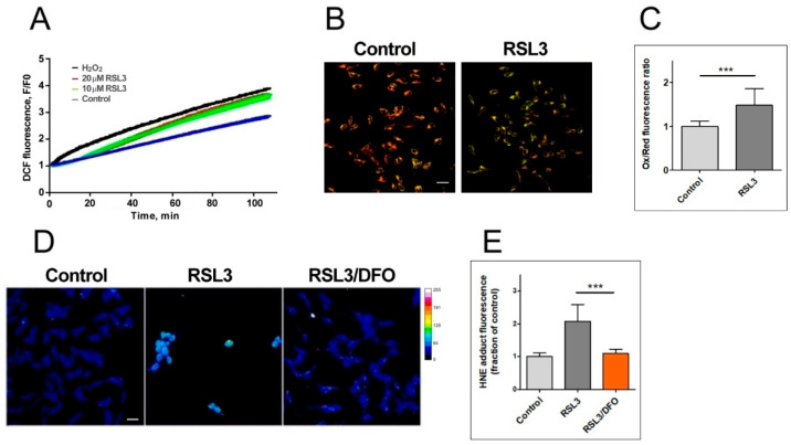
To assess the ferroptotic characteristics of RSL3-treated cells, we determined markers of ROS production and membrane oxidation (Figure 3).
为了评估 RSL3 处理的细胞的铁死亡特征,我们确定了 ROS 产生和膜氧化的标记(图 3 )。
Figure 3. 图 3.
RSL3 induces an increase in ROS production and lipid peroxidation. (A) SH-SY5Y cells were loaded with H2DCFDA and were then treated at t = 0 with 10 µM or 20 µM RSL3 or 250 µM H2O2; DCF fluorescence intensity was recorded over time. Values represent means of 5 replicates per experimental condition. N = 3 independent experiments. (B) SH-SY5Y cells, treated for 3 h with 5 µM RSL3 or vehicle, were stained with BODIPY C11. Representative images of the overlapping values collected by the red (reduced, 590 nm emission) and green (oxidized, 510 nm emission) fluorescence channels for each condition are shown. Scale bar 20 µm. (C) Quantification of the ratio of the green to red fluorescence intensity. Values represent mean ± SD of 150 cells; *** p < 0.001. (D) Cells were treated for 4 h without (control) or with 5 µM RSL3. Representative frames of HNE adducts immunofluorescence are shown. Scale bar 20 µm. The upper right corner shows a thermal fluorescence intensity scale. (E) Quantification of HNE fluorescence intensity. Values represent mean ± SD for 100–120 cells per experimental condition, N = 3 independent experiments, *** p < 0.001.
RSL3 诱导 ROS 产生和脂质过氧化增加。 ( A ) SH-SY5Y 细胞加载 H2DCFDA,然后在 t = 0 时用 10 µM 或 20 µM RSL3 或 250 µM H 2 O 2处理;随着时间的推移记录 DCF 荧光强度。值代表每个实验条件 5 次重复的平均值。 N = 3 个独立实验。 ( B ) 用 5 µM RSL3 或媒介物处理 3 小时的 SH-SY5Y 细胞,用 BODIPY C11 染色。显示了每种条件下红色(还原,590 nm 发射)和绿色(氧化,510 nm 发射)荧光通道收集的重叠值的代表性图像。比例尺 20 µm。 ( C ) 绿色荧光强度与红色荧光强度之比的量化。数值代表 150 个细胞的平均值±SD; *** p < 0.001。 ( D ) 细胞在不使用(对照)或使用 5 µM RSL3 的情况下处理 4 小时。显示了 HNE 加合物免疫荧光的代表性框架。比例尺 20 µm。右上角显示热荧光强度刻度。 ( E ) HNE 荧光强度的量化。值代表每个实验条件下 100-120 个细胞的平均值 ± SD,N = 3 个独立实验,*** p < 0.001。
The generation of ROS was evaluated via DCF fluorescence. The addition of 20 µM RSL3 induced a robust ROS production, similar to that elicited by 250 µM H2O2 (Figure 3A). We then evaluated lipid peroxidation levels after 3 h of RSL3 treatment using the BODIPY C11 probe, which is a long-chain oxidizable molecule that inserts into cell membranes. In the reduced state, this probe emits red fluorescence (λ emission 590 nm); however, when oxidized, its emission/excitation pattern changes towards green fluorescence (λ emission 510 nm). We found that RSL3 treatment for 3 h produced a significant increase in the ratio of green fluorescence to red fluorescence compared to the control, indicative of lipid peroxidation (Figure 3B,C). Lipid peroxidation was further assessed by determining the formation of HNE-protein adducts; 4-HNE is a highly reactive aldehyde resulting from the lipid peroxidation of polyunsaturated fatty acids such as linoleic and arachidonic. We found that treatment with RSL3 increased the formation of protein-HNE adducts, and that co-incubation with the iron chelator deferoxamine (DFO) circumvented this increase (Figure 3D,E).
通过 DCF 荧光评估 ROS 的产生。添加 20 µM RSL3 诱导了强劲的 ROS 产生,类似于 250 µM H 2 O 2引发的情况(图 3 A)。然后,我们使用 BODIPY C11 探针(一种插入细胞膜的长链可氧化分子)评估了 RSL3 处理 3 小时后的脂质过氧化水平。在还原状态下,该探针发出红色荧光(发射波长590 nm);然而,当被氧化时,其发射/激发模式会变为绿色荧光(发射波长 510 nm)。我们发现,与对照相比,RSL3 处理 3 小时后绿色荧光与红色荧光的比率显着增加,表明脂质过氧化(图 3 B、C)。通过测定 HNE-蛋白质加合物的形成进一步评估脂质过氧化作用; 4-HNE 是一种高活性醛,由亚油酸和花生四烯酸等多不饱和脂肪酸的脂质过氧化作用产生。我们发现用 RSL3 处理增加了蛋白质-HNE 加合物的形成,并且与铁螯合剂去铁胺 (DFO) 共孵育避免了这种增加(图 3 D、E)。
3.1.3. RSL3 Induces Non-Apoptotic Cell Death in SH-SY5Y Neuroblastoma Cells
3.1.3. RSL3 诱导 SH-SY5Y 神经母细胞瘤细胞非凋亡性细胞死亡
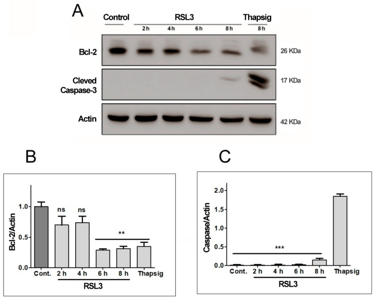
To ascertain the non-apoptotic nature of RSL3-mediated SH-SY5Y cell death, the levels of two apoptosis-related proteins, Bcl-2 and cleaved Caspase-3, were determined (Figure 4).
为了确定RSL3介导的SH-SY5Y细胞死亡的非凋亡性质,测定了两种凋亡相关蛋白Bcl-2和裂解的Caspase-3的水平(图4 )。
Figure 4. 图 4.
RSL3 decreases Bcl-2 levels but does not induce Caspase-3 cleavage. Extracts of cells treated with 5 µM RSL3 for different times, or with 5 µM thapsigargin (Thapsig) for 8 h, were analyzed by Western blot to evaluate Bcl-2 and cleaved Caspase-3 levels. Actin was used as a loading control. (A) Image of a representative blot. (B) Densitometric quantification of Bcl-2/Actin levels normalized to control. (C) Densitometric quantification of cleaved Caspase-3/Actin levels normalized to control. after treatment with RSL3 for different times, or with 5 µM thapsigargin for 16 h. Mean ± SEM values are shown. N = 3 independent experiments; ns = not significant, ** p < 0.01, *** p < 0.001.
RSL3 降低 Bcl-2 水平,但不诱导 Caspase-3 裂解。用 5 µM RSL3 处理不同时间或用 5 µM 毒胡萝卜素 (Thhapsig) 处理 8 小时的细胞提取物通过蛋白质印迹进行分析,以评估 Bcl-2 和裂解的 Caspase-3 水平。肌动蛋白用作加载对照。 ( A ) 代表性印迹的图像。 ( B ) Bcl-2/肌动蛋白水平的光密度定量标准化为对照。 ( C ) 裂解的 Caspase-3/肌动蛋白水平的光密度定量标准化为对照。用 RSL3 处理不同时间,或用 5 µM 毒胡萝卜素处理 16 小时后。显示平均值±SEM 值。 N = 3 个独立实验; ns = 不显着,** p < 0.01,*** p < 0.001。
We found that RSL3 treatment for 6 h or 8 h significantly decreased Bcl-2 protein levels (Figure 4A,B), whereas Caspase-3 was detectable at very low levels only after 8 h of treatment with RSL3 (Figure 4A). Treatment with thapsigargin for 16 h served as a positive control for the induction of apoptosis, allowing comparison of the levels of cleaved caspase-3 in comparison to those induced by RSL3 treatment for 8 h. The levels of cleaved caspase-3 were 12–14-fold higher after treatment with thapsigargin, as compared to RSL3 treatment for 8 h (Figure 4C). Based on the cleaved caspase-3 levels, it appears that in SH-SY5Y cells, RSL3 induces mostly non-apoptotic cell death, with the caveat that at longer times of incubation (6–8 h), apoptosis markers are observed (decreased Bcl-2 protein levels).
我们发现,RSL3 处理 6 小时或 8 小时显着降低了 Bcl-2 蛋白水平(图 4 A、B),而仅在 RSL3 处理 8 小时后才可检测到非常低的水平的 Caspase-3(图 4 A)。用毒胡萝卜素处理 16 小时作为诱导细胞凋亡的阳性对照,从而可以将裂解的 caspase-3 水平与 RSL3 处理 8 小时诱导的水平进行比较。与 RSL3 处理 8 小时相比,用毒胡萝卜素处理后,裂解的 caspase-3 水平高出 12-14 倍(图 4 C)。根据裂解的 caspase-3 水平,似乎在 SH-SY5Y 细胞中,RSL3 诱导大部分非凋亡性细胞死亡,但需要注意的是,在较长的孵育时间(6-8 小时)下,观察到凋亡标记物(Bcl 减少) -2蛋白质水平)。
Overall, the data substantiate the notion that SH-SY5Y cells treated with RSL3 is a validated model for the study of ferroptosis.
总体而言,数据证实了用 RSL3 处理的 SH-SY5Y 细胞是铁死亡研究的有效模型的观点。
3.2. Treatment with RSL3 Causes a Delayed Increase in Intracellular Calcium Concentration
3.2. RSL3 治疗导致细胞内钙浓度延迟增加
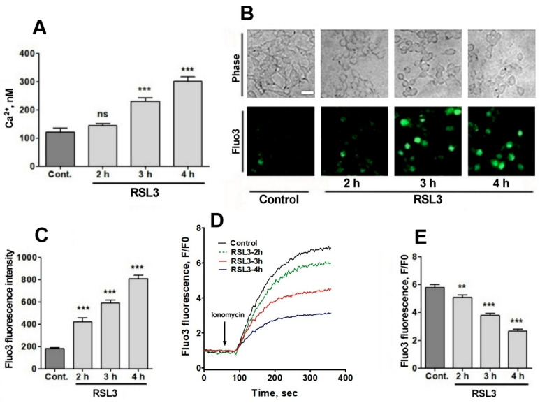
Since RSL3 induced an increase in ROS, plus the fact that the intracellular calcium channels RyR and IP3R are activated by oxidation [23,24,25,26], the possible effects of RSL3 on increasing the intracellular calcium concentration in SH-SY5Y cells were investigated. To measure the intracellular calcium concentration, we used fura-2, a ratiometric calcium probe that has different fluorescence absorption in its calcium-bound or -unbound states [33]. Calcium-free fura-2 has an excitation maximum at 340 nm, whereas when bound to calcium, this maximum shifts to 380 nm. Therefore, it is possible to normalize the calcium concentrations with respect to the amount of loaded probe, exciting at the two excitation wavelengths and recording at 510 nm. Preliminary studies did not detect RSL3-induced increases in intracellular calcium at short (0–20 min) time periods, which is in line with the calcium dynamics observed in RSL3-induced ferroptosis in primary hippocampal neurons [27]. To assess whether calcium concentrations were altered at longer (hours) times, cells were treated with RSL3 for different times and then loaded with fura-2 (Figure 5A).
由于RSL3诱导ROS增加,加上细胞内钙通道RyR和IP 3 R被氧化激活的事实[ 23,24,25,26 ],RSL3对增加SH - SY5Y细胞内钙浓度的可能影响细胞进行了研究。为了测量细胞内钙浓度,我们使用了 fura-2,这是一种比例钙探针,在钙结合或非结合状态下具有不同的荧光吸收[ 33 ]。无钙的 fura-2 在 340 nm 处具有激发最大值,而当与钙结合时,该最大值移动到 380 nm。因此,可以根据负载探针的量标准化钙浓度,在两个激发波长下激发并在 510 nm 处记录。初步研究并未检测到 RSL3 在短时间内(0-20 分钟)诱导的细胞内钙增加,这与在 RSL3 诱导的原代海马神经元铁死亡中观察到的钙动态一致 [ 27 ]。为了评估钙浓度是否在较长(小时)时间内发生变化,用 RSL3 处理细胞不同时间,然后加载 fura-2(图 5 A)。
Figure 5. 图 5.
RSL3 alters calcium concentrations after a delay period. (A) Cells pre-treated with 5 µM RSL3 for different times were loaded with fura-2 and fluorescence was recorded to subsequently determine cytoplasmic calcium concentrations. Values represent mean ± SEM of a representative experiment from 3 independent experiments with 3 replicates per experimental condition. Differences were determined by one-way ANOVA followed by Dunnett’s post hoc test. (B) Cells pre-treated with 5 µM RSL3 or vehicle (DMSO, control) were loaded with fluo-3 and fluorescence was recorded. Representative images of each treatment are shown. Scale bar 20 µm. (C) Quantification of cell fluorescence after the different treatments. Values are given as mean ± SEM; 40 cells per condition. (D) Plots of the ratio of fluorescence intensity over initial fluorescence (F/F0) as a function of time, before and after the addition of ionomycin (arrow). (E) Maximum F/F0 ratio reached after the addition of ionomycin for each condition. Mean ± SEM of the last three points for each experimental condition with points in triplicate. ns = not significant; ** p < 0.01; *** p < 0.001.
RSL3 在延迟期后改变钙浓度。 ( A ) 用 5 µM RSL3 预处理不同时间的细胞加载 fura-2,并记录荧光以随后测定细胞质钙浓度。值代表来自 3 个独立实验的代表性实验的平均值±SEM,每个实验条件重复 3 次。通过单向方差分析和 Dunnett 事后检验确定差异。 ( B ) 用 5 µM RSL3 或载体(DMSO,对照)预处理的细胞加载 Fluo-3 并记录荧光。显示了每种治疗的代表性图像。比例尺 20 µm。 ( C ) 不同处理后细胞荧光的定量。数值以平均值±SEM的形式给出;每个条件 40 个细胞。 ( D ) 在添加离子霉素之前和之后,荧光强度与初始荧光的比率 (F/F0) 作为时间的函数的图(箭头)。 ( E ) 在每种条件下添加离子霉素后达到的最大 F/F0 比率。每个实验条件下最后三个点的平均值±SEM,点一式三份。 ns = 不显着; ** p < 0.01; *** p < 0.001。
A gradual increase in cytoplasmic calcium concentration was found in cells treated with 5 µM RSL3, from about 100 nM in the untreated situation to 300 nM after 4 h of RSL3 treatment, with significant differences at 3 h and 4 h post RSL3 addition (Figure 5A). The RSL3-induced cytoplasmic calcium increase was further explored by treating cells with RSL3 for different times and then loading them with fluo-3, a calcium sensor with higher Kd than fura-2 [33]. The fluorescence intensity of fluo-3 increased in proportion to the duration of the previous RSL3 treatment (Figure 5B,C).
在用 5 µM RSL3 处理的细胞中发现细胞质钙浓度逐渐增加,从未处理情况下的约 100 nM 增加到 RSL3 处理 4 小时后的 300 nM,添加 RSL3 后 3 小时和 4 小时有显着差异(图 5)一个)。通过用 RSL3 处理细胞不同时间,然后加载 Fluo-3(一种 Kd 高于 fura-2 的钙传感器),进一步探讨了 RSL3 诱导的细胞质钙增加[ 33 ]。 Fluo-3 的荧光强度与先前 RSL3 处理的持续时间成比例增加(图 5 B、C)。
To further typify the RSL3-induced intracellular calcium upsurge, cells were treated for different times with RSL3 and then challenged with the calcium ionophore ionomycin to allow for calcium influx from the extracellular medium. It was found that fluo-3 responded to ionomycin with increased fluorescence (Figure 5D,E). In addition, it was observed that the response was inversely related to the pre-incubation time with RSL3, a result consistent with an increased intracellular calcium concentration as a function of increased times of RSL3 treatment.
为了进一步表征RSL3诱导的细胞内钙离子激增,用RSL3处理细胞不同时间,然后用钙离子载体离子霉素激发,以允许钙从细胞外介质流入。结果发现,fluo-3 对离子霉素的反应荧光增强(图 5 D、E)。此外,观察到反应与RSL3的预孵育时间成反比,该结果与作为RSL3处理时间增加的函数的细胞内钙浓度增加一致。
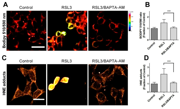
Due to the relevance of lipid peroxidation in ferroptosis, a possible causal relationship between RSL3-induced calcium upsurge and lipid peroxidation was investigated. To this end, we determined the effects of BAPTA-AM on the RSL3-induced oxidation of BODIPY C11. As previously carried out, we also measured the formation of 4-HNE-protein adducts after RSL3 treatment.
由于铁死亡中脂质过氧化的相关性,研究了RSL3诱导的钙离子激增与脂质过氧化之间可能的因果关系。为此,我们确定了 BAPTA-AM 对 RSL3 诱导的 BODIPY C11 氧化的影响。如前所述,我们还测量了 RSL3 处理后 4-HNE-蛋白加合物的形成。
We found that pre-incubation with BAPTA-AM largely prevented RSL3-induced BODIPY oxidation (Figure 6A,B). Likewise, we found that the levels of 4-HNE adducts were significantly lower in BAPTA-AM-treated cells (Figure 6C,D). Thus, it is apparent that mechanistically, RSL3-induced intracellular calcium increase precedes lipid peroxidation.
我们发现,与 BAPTA-AM 预孵育很大程度上阻止了 RSL3 诱导的 BODIPY 氧化(图 6 A、B)。同样,我们发现 BAPTA-AM 处理的细胞中 4-HNE 加合物的水平显着降低(图 6 C、D)。因此,很明显,从机制上讲,RSL3诱导的细胞内钙增加先于脂质过氧化。
Figure 6. 图 6.
Calcium chelation protects cells against RSL3-induced lipid peroxidation. Cells were pre-incubated for 1 h with 5 µM BAPTA-AM followed by co-incubation for 3 h with 5 µM RSL3 and analysis of lipid peroxidation with either BODIPY C11 or anti-HNE immunofluorescence as described in Methods. (A) Representative images of BODIPY fluorescence upon treatment with RSL3, without (control) or after pre-treatment with BAPTA-AM. Scale bar 20 µm. (B) Quantification of the ratio of fluorescence intensity of the oxidized (green) channel to that of the reduced (red) channel. At least 80 cells per condition were quantified from N = 3 independent experiments. Mean ± SD values are shown. (C) Representative images of immunodetection against 4-HNE adducts. Scale bar 15 µm. (D) Quantification of fluorescence intensity. Values represent mean ± SD from 75–90 cells quantified for each experimental condition from N = 3 independent experiments. Both in (B,D) differences were evaluated by one-way ANOVA, followed by Dunnett’s post hoc test. *** p < 0.001.
钙螯合可保护细胞免受 RSL3 诱导的脂质过氧化作用。将细胞与 5 µM BAPTA-AM 预孵育 1 小时,然后与 5 µM RSL3 共孵育 3 小时,并使用 BODIPY C11 或抗 HNE 免疫荧光分析脂质过氧化,如方法中所述。 ( A ) 用 RSL3 处理、未用 BAPTA-AM(对照)或用 BAPTA-AM 预处理后的 BODIPY 荧光的代表性图像。比例尺 20 µm。 ( B ) 氧化(绿色)通道的荧光强度与还原(红色)通道的荧光强度之比的定量。从 N = 3 次独立实验中对每种条件至少 80 个细胞进行定量。显示平均值±SD 值。 ( C ) 针对 4-HNE 加合物的免疫检测的代表性图像。比例尺 15 µm。 ( D ) 荧光强度的定量。值代表来自 N = 3 个独立实验的每个实验条件下量化的 75-90 个细胞的平均值±SD。 ( B , D ) 中的差异均通过单向方差分析进行评估,然后进行 Dunnett 事后检验。 *** p < 0.001。
3.3. The IP3R Calcium Channel Mediates RSL3-Induced Increase in Intracellular Calcium
3.3. IP 3 R 钙通道介导 RSL3 诱导的细胞内钙增加
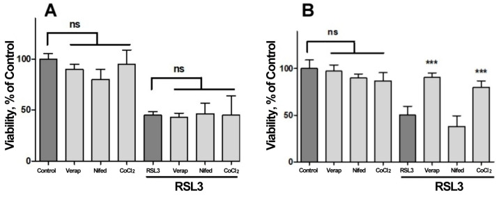
We next investigated the possible participation of calcium channels on the RSL3-induced increase in intracellular calcium concentration (Figure 7). Veparamil and Nifedipine, two L-type voltage-gated calcium channel blockers [36,37], and CoCl2, a nonspecific calcium channel antagonist, did not afford protection against RSL3 treatment (Figure 7A). In another series of experiments, the extracellular calcium chelator EGTA and the cell-permeant calcium chelator BAPTA-AM were tested as possible protectors of RSL3 toxicity. In these series, the addition of EGTA to the culture medium failed to diminish the effects on viability due to RSL3 treatment (Figure 7B). In contrast, loading cells with BAPTA-AM prior to RSL3 treatment significantly protected SH-SY5Y cell viability (Figure 7B). As a positive control, protection by DFO was also observed (Figure 7B).
接下来我们研究了钙通道对RSL3诱导的细胞内钙浓度增加的可能参与(图7 )。 Veparamil 和 Nifedipine,两种 L 型电压门控钙通道阻滞剂 [ 36 , 37 ],以及 CoCl 2 ,一种非特异性钙通道拮抗剂,不能提供针对 RSL3 治疗的保护作用(图 7 A)。在另一系列实验中,测试了细胞外钙螯合剂 EGTA 和细胞渗透性钙螯合剂 BAPTA-AM 作为 RSL3 毒性的可能保护剂。在这些系列中,在培养基中添加 EGTA 未能减少 RSL3 处理对活力的影响(图 7 B)。相比之下,在 RSL3 处理之前用 BAPTA-AM 加载细胞可显着保护 SH-SY5Y 细胞活力(图 7 B)。作为阳性对照,还观察到 DFO 的保护作用(图 7 B)。
Figure 7. 图 7.
Both BAPTA and DFO protect against RSL3-induced decrease in cell viability. Cells were incubated for 90 min with 300 µM Verapamil (Verap), 5 µM Nifedipine (Nifed) or 100 µM CoCl2 (A) or with 25 µM DFO, 2.5 mM EGTA or 2.5 µM BAPTA-AM (B), followed by incubation for 4 h with 5 µM RSL3. After that, cell viability was estimated by the MTT assay. Values represent mean ± SEM. N = 3 independent experiments with determinations done in sextuplicate. Differences were assessed by one-way ANOVA followed by Dunnett’s test. *** p < 0.001 compared to RSL3 alone treatment; ns = not significant.
BAPTA 和 DFO 均可防止 RSL3 诱导的细胞活力下降。将细胞与 300 µM 维拉帕米 (Verap)、5 µM 硝苯地平 (Nifed) 或 100 µM CoCl 2 ( A ) 或与 25 µM DFO、2.5 mM EGTA 或 2.5 µM BAPTA-AM ( B ) 一起孵育 90 分钟,然后孵育使用 5 µM RSL3 孵育 4 小时。之后,通过 MTT 测定评估细胞活力。值代表平均值±SEM。 N = 3 个独立实验,测定一式六份。通过单向方差分析和 Dunnett 检验评估差异。 *** 与单独 RSL3 治疗相比, p < 0.001; ns = 不显着。
Since viability was not protected by compounds that hinder calcium influx from the extracellular medium, it is likely that the observed calcium increases induced by RSL3 treatment depend on calcium release from intracellular calcium stores. Considering that (i) the endoplasmic reticulum (ER) is the most important store of intracellular calcium, (ii) the release of calcium from this organelle is mainly regulated by two channels, the RyR and the IP3R channels, and (iii) that the intracellular calcium channels RyR and IP3R are activated by oxidation [23,24,25,26], the putative mediation by these channels in cell death induced by RSL3 in SH-SY5Y cells was evaluated (Figure 8).
由于细胞活力不受阻碍钙从细胞外培养基流入的化合物的保护,因此观察到的由RSL3处理诱导的钙增加可能取决于细胞内钙储存的钙释放。考虑到 (i) 内质网 (ER) 是细胞内钙最重要的储存库,(ii) 该细胞器的钙释放主要由 RyR 和 IP 3 R 通道这两个通道调节,以及 (iii)细胞内钙通道 RyR 和 IP 3 R 通过氧化被激活[23,24,25,26 ] ,这些通道在由评估了SH-SY5Y细胞中的RSL3(图8 )。
Figure 8. 图 8.
The role of IP3R channels in RSL3-induced ferroptosis. (A) SH-SY5Y cells were pre-incubated for 1 h with 100 µM Ryanodine, 10 µM xestospongin B (XeB) or 1 mM carbachol (Cch) and were then incubated for 4 h with RSL3. Viability was determined by the MTT assay; ns = not significant, *** p < 0.001. (B) Representative Western blots against IP3R1 after Cch treatments for 0, 1, 4 or 6 h. (C) Quantification of relative protein levels of IP3R1 after Cch treatment. Values represent the mean ± SD from N = 3 independent experiments; *** p < 0.001 compared to Control.
IP 3 R 通道在 RSL3 诱导的铁死亡中的作用。 ( A ) SH-SY5Y 细胞与 100 µM Ryanodine、10 µM xestospongin B (XeB) 或 1 mM carbachol (Cch) 预孵育 1 小时,然后与 RSL3 一起孵育 4 小时。通过MTT测定测定活力; ns = 不显着,*** p < 0.001。 ( B ) Cch 处理 0、1、4 或 6 小时后针对 IP 3 R1 的代表性蛋白质印迹。 ( C ) Cch 处理后 IP 3 R1 相对蛋白水平的定量。数值代表 N = 3 个独立实验的平均值±SD; *** 与对照相比, p < 0.001。
No effects on RSL3-mediated cell death were observed when cells were pre-incubated for 1 h with 100 µM ryanodine, a specific RyR channel inhibitor at µM concentrations (Figure 8A). This result agrees with the fact that the SH-SY5Y cell line used in this work does not express RyR channels (Supplementary Figure S1). In contrast, treatment with xestospongin B, a powerful and specific IP3R channel blocker, protected cells from RSL3-induced cell death (Figure 8A). The participation of the IP3R channel in this process was further evaluated with carbachol, a cholinergic agonist that reduces IP3R protein levels in SH-SY5Y cells [38]. Pre-incubation with Cch effectively produced a time-dependent decrease in IP3R1 levels (Figure 8B,C) and protected cells from RSL3-induced cell death (Figure 8A). Overall, these results indicate that RSL3 induces an increase in intracellular calcium that is mediated, in large part, by IP3R channels, and that this increase is germane to RSL3-induced ferroptotic cell death.
当细胞与 100 µM ryanodine(一种 µM 浓度的特定 RyR 通道抑制剂)预孵育 1 小时时,未观察到对 RSL3 介导的细胞死亡有影响(图 8 A)。该结果与本工作中使用的 SH-SY5Y 细胞系不表达 RyR 通道的事实一致(补充图 S1 )。相比之下,用 xestospongin B(一种强效且特异性的 IP 3 R 通道阻断剂)治疗可保护细胞免遭 RSL3 诱导的细胞死亡(图 8 A)。使用卡巴胆碱(一种胆碱能激动剂,可降低 SH-SY5Y 细胞中 IP 3 R 蛋白水平)进一步评估 IP 3 R 通道在此过程中的参与[ 38 ]。与 Cch 预孵育有效地产生了 IP 3 R1 水平的时间依赖性下降(图 8 B、C),并保护细胞免遭 RSL3 诱导的细胞死亡(图 8 A)。总的来说,这些结果表明RSL3诱导细胞内钙的增加,这在很大程度上是由IP 3 R通道介导的,并且这种增加与RSL3诱导的铁死亡细胞死亡密切相关。
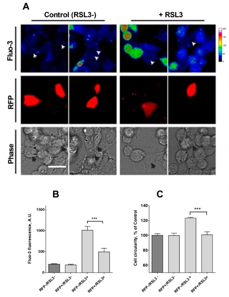
To further confirm the participation of IP3R channels in the calcium increase observed in response to RSL3 treatment, the expression of IP3R1, the main isoform expressed in SH-SY5Y cells [39], was downregulated. To this end, cells were transiently transfected with a plasmid containing a shRNA sequence against IP3R1 tagged to a RFP sequence. Transfected cells were treated, or not, with RSL3 and then loaded with fluo-3 to analyze cytoplasmic calcium levels. Fluo-3 fluorescence intensity was determined in RFP-positive (RFP+) cells, which expressed the interfering sequence and RFP-negative cells (RFP-), that did not express the shRNA sequence.
为了进一步确认 IP 3 R 通道参与 RSL3 处理后观察到的钙增加,SH-SY5Y 细胞中表达的主要异构体 IP 3 R1 的表达下调[ 39 ]。为此,用含有针对标记有 RFP 序列的 IP 3 R1 的 shRNA 序列的质粒瞬时转染细胞。转染的细胞用RSL3处理或不处理,然后加载fluo-3以分析细胞质钙水平。在表达干扰序列的 RFP 阳性 (RFP+) 细胞和不表达 shRNA 序列的 RFP 阴性细胞 (RFP-) 中测定 Fluo-3 荧光强度。
Under control conditions, no differences between RFP+ and RFP- cells were found, with a consistently low fluo-3 signal. When both groups were compared after treatment with RSL3, the fluo-3 fluorescence intensity was significantly lower in cells with knockdown for IP3R1 (Figure 9B), an indication that IP3R1 mediates the RSL3-induced calcium upsurge.
在对照条件下,未发现 RFP+ 和 RFP- 细胞之间存在差异,且 Fluo-3 信号始终较低。当用 RSL3 处理后比较两组时,在 IP 3 R1 敲低的细胞中,fluo-3 荧光强度显着较低(图 9B ),表明 IP 3 R1 介导 RSL3 诱导的钙离子激增。
Figure 9. 图 9.
Knockdown of IP3R1 reduces the increase in intracellular calcium concentration induced by RSL3. Cells previously transfected with a shRNA construct against IP3R1 with a RFP tag (RFP+) were treated for 4 h with 5 µM RSL3 or vehicle, then loaded with fluo-3 and frames were obtained with red and green fluorescence filters. (A) Representative images in thermal scale of non-treated cells (columns 1 and 2) and RSL3-treated cells (columns 3 and 4). Scale bar 20 µm. White (Fluo-3) and black (Phase) arrowheads mark the position of RFP-positive cells. (B) Quantification of fluo-3 fluorescence in RFP+ cells, which corresponds to IP3R1 shRNA transfected cells, and RFP- cells, which do not express the IP3R1 construct, without (RSL3-) or with (RSL3+) RSL3 treatment. (C) Estimation of roundness in RFP+ and RFP- cells treated or not with RSL3. Values represent mean ± SEM. Between 32 and 115 cells were evaluated per experimental condition from 2 independent experiments. *** p = 0.001 comparing the RFP-/RSL3+ and the RFP-/RSL3+ conditions. No significant changes were detected between the RFP-/RSL3-, RFP+/RSL3- and RFP+/RSL3+ conditions.
IP 3 R1 的敲低可降低 RSL3 诱导的细胞内钙浓度的增加。先前用带有 RFP 标签 (RFP+) 的针对 IP 3 R1 的 shRNA 构建体转染的细胞用 5 µM RSL3 或载体处理 4 小时,然后加载 Fluo-3,并使用红色和绿色荧光滤光片获得框架。 ( A ) 未处理细胞(第 1 列和第 2 列)和 RSL3 处理细胞(第 3 列和第 4 列)的热标度代表性图像。比例尺 20 µm。白色 (Fluo-3) 和黑色 (Phase) 箭头标记 RFP 阳性细胞的位置。 ( B ) RFP+ 细胞(对应于 IP 3 R1 shRNA 转染细胞)和 RFP- 细胞(不表达 IP 3 R1 构建体)中 Fluo-3 荧光的定量,无需 (RSL3-) 或经过 (RSL3+) RSL3 处理。 ( C ) 估计用 RSL3 处理或未处理的 RFP+ 和 RFP- 细胞的圆度。值代表平均值±SEM。在 2 个独立实验中,每个实验条件评估了 32 至 115 个细胞。 *** 比较 RFP-/RSL3+ 和 RFP-/RSL3+ 条件时, p = 0.001。 RFP-/RSL3-、RFP+/RSL3- 和 RFP+/RSL3+ 条件之间未检测到显着变化。
Because of a low efficiency (5–7%) of transfection with the shRNA sequence against IP3R1, it was not possible to evaluate RSL3-induced ferroptotic death in IP3R1 shRNA-transfected cells using the MTT assay, which was used to generate the results described in Figure 1. As an alternative, RSL3-induced roundness, a bona fide ferroptosis indicator, was evaluated (Figure 9C). The data indicated that IP3R1 shRNA-transfected cells were significantly more resistant to the change towards roundness induced by RSL3 than non-transfected cells. IP3R1 shRNA-transfected cells were also resistant to the RSL3-induced changes in area and perimeter described in Figure 2 (Supplementary Figure S2). Overall, the data strongly suggest that IP3R1 mediates the ferroptotic events induced by RSL3.
由于针对 IP 3 R1 的 shRNA 序列转染效率较低 (5-7%),因此不可能使用 MTT 测定来评估 RSL3 诱导的 IP 3 R1 shRNA 转染细胞中的铁死亡。生成如图 1所示的结果。作为替代方案,评估了 RSL3 诱导的圆度(一种真正的铁死亡指标)(图 9C )。数据表明IP 3 R1 shRNA转染的细胞比未转染的细胞对RSL3诱导的圆度变化的抵抗力明显更强。 IP 3 R1 shRNA 转染细胞也能抵抗 RSL3 诱导的面积和周长变化,如图 2所示(补充图 S2 )。总体而言,数据强烈表明 IP 3 R1 介导 RSL3 诱导的铁死亡事件。
3.4. RSL Treatment Induces an Increase in Mitochondrial Calcium
3.4. RSL 治疗可导致线粒体钙增加
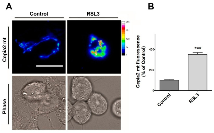
Since through mitochondria-associated ER membranes (MAMs), IP3R participates in calcium entry into mitochondria [40], the relative levels of mitochondrial calcium after treatment with RSL3 were investigated using the genetically encoded mitochondria calcium probe CEPIA2mt (Figure 10).
由于IP 3 R通过线粒体相关的ER膜(MAM)参与钙进入线粒体[ 40 ],因此使用基因编码的线粒体钙探针CEPIA2mt研究了RSL3处理后线粒体钙的相对水平(图10 )。
Figure 10. 图 10.
RSL3 induces an increase in mitochondrial calcium levels. Cells transfected with the mitochondrial calcium sensor CEPIA2mt were treated with 5 µM RSL3 for 4 h or with control solution, and the fluorescence intensity was recorded. (A) Representative images of both conditions shown in thermal scale (right-hand bar). Size bar 30 µm. (B) Quantification of fluorescence intensity. Values represent Mean ± SEM from 58–62 cells quantified for each experimental condition. Difference between mean values was determined by unpaired two-tail t-test. N = 2 independent experiments. *** p < 0.001.
RSL3 诱导线粒体钙水平增加。用线粒体钙传感器 CEPIA2mt 转染的细胞用 5 µM RSL3 或对照溶液处理 4 小时,并记录荧光强度。 ( A ) 以热标度显示两种情况的代表性图像(右侧条)。尺寸条 30 µm。 ( B ) 荧光强度的定量。值代表每个实验条件下量化的 58-62 个细胞的平均值±SEM。平均值之间的差异通过不配对的双尾t检验确定。 N = 2 个独立实验。 *** p < 0.001。
Cells treated with RSL3 showed a striking 3.5-fold increase in mitochondrial calcium levels (Figure 10A,B). Overall, these results indicate that RSL3-induced ferroptosis in SH-SY5Y cells provokes an increase in cytoplasmic and mitochondrial calcium levels that are mediated by the IP3R channel.
用 RSL3 处理的细胞显示线粒体钙水平显着增加 3.5 倍(图 10 A、B)。总体而言,这些结果表明,SH-SY5Y 细胞中 RSL3 诱导的铁死亡会引起由 IP 3 R 通道介导的细胞质和线粒体钙水平增加。
4. Discussion 4. 讨论
The participation of calcium in ferroptosis is a matter of debate. Initial studies using HT-1080 epithelial cells discarded a role for calcium under the observation that ferroptosis was not blocked by the extracellular calcium chelator EGTA or by the intracellular chelator BAPTA-AM [2]. Nonetheless, other reports proposed that ferroptosis and oxytosis are the same phenomenon [20,21]. Because oxytosis is highly dependent on calcium entry into the cell, here, we evaluated the participation of calcium in a model of RSL3-induced ferroptosis using the dopaminergic cell line SH-SY5Y. Of note, this is the first report that establishes a firm link between calcium and ferroptosis in this neuroblastoma cell line. In addition to previous works, which reported that plasma membrane calcium channels [21,41] or RyR channels [27,41] act as mediators in the increase in intracellular calcium induced by RSL3, here, we additionally identified IP3R channels as mediators of the calcium increase induced by RSL3.
钙在铁死亡中的参与是一个有争议的问题。使用 HT-1080 上皮细胞的初步研究放弃了钙的作用,观察结果是细胞外钙螯合剂 EGTA 或细胞内螯合剂 BAPTA-AM 不会阻断铁死亡 [ 2 ]。尽管如此,其他报告提出铁死亡和氧化中毒是同一现象[ 20 , 21 ]。因为催氧作用高度依赖于钙进入细胞,所以在这里,我们使用多巴胺能细胞系 SH-SY5Y 评估了钙在 RSL3 诱导的铁死亡模型中的参与。值得注意的是,这是第一份在神经母细胞瘤细胞系中建立钙和铁死亡之间牢固联系的报告。除了先前报道质膜钙通道 [ 21 , 41 ] 或 RyR 通道 [ 27 , 41 ] 在 RSL3 诱导的细胞内钙增加中充当介质的工作之外,在这里,我们还确定了 IP 3 R 通道作为介质RSL3 诱导的钙增加。
We found that RSL3 affected the viability of SH-SY5Y cells in a dose- and time-dependent manner. Thus, RSL3 induced 50% cell death at times as short as 4 h. Treatment with RSL3 also induced landmarks of ferroptosis, such as an increase in ROS levels and a remarkable change in cell morphology, with a significant increase in cell roundness. Arguably, the changes in ROS production and cell morphology are a preamble to cell death. Albeit the direct inhibitory effects of RSL3 as an inhibitor of GPx4 have been recently questioned based on studies performed in cell-free systems [42], our results support the role of RSL3 as an effective ferroptosis-inducing agent.
我们发现RSL3以剂量和时间依赖性方式影响SH-SY5Y细胞的活力。因此,RSL3 在短短 4 小时内即可诱导 50% 的细胞死亡。 RSL3处理还诱导了铁死亡的标志,例如ROS水平的增加和细胞形态的显着变化,以及细胞圆度的显着增加。可以说,ROS 产生和细胞形态的变化是细胞死亡的前奏。尽管基于无细胞系统中进行的研究,RSL3 作为 GPx4 抑制剂的直接抑制作用最近受到质疑 [ 42 ],但我们的结果支持 RSL3 作为有效的铁死亡诱导剂的作用。
Associated with ROS production, there was an increase in membrane lipid peroxidation, a hallmark of ferroptosis. In accordance with the participation of iron in ferroptosis, co-incubation with the iron chelator DFO precluded lipid peroxidation, as detected by the levels of 4-HNE-protein adducts. Overall, these results indicate that RSL3 induces a robust ferroptotic response in SH-SY5Y cells.
与 ROS 产生相关的是膜脂过氧化的增加,这是铁死亡的标志。根据铁在铁死亡中的参与,与铁螯合剂 DFO 共孵育可防止脂质过氧化,如通过 4-HNE-蛋白质加合物的水平检测到的。总体而言,这些结果表明 RSL3 在 SH-SY5Y 细胞中诱导强烈的铁死亡反应。
In consideration of a putative participation of calcium in ferroptosis, we used fluorescent probes to detect possible alterations in calcium levels produced by RSL3. Firstly, we observed that the application of RSL3 did not trigger an immediate (seconds–minutes) increase in intracellular calcium concentration in the way that, for example, thapsigargin, carbachol, or even hydrogen peroxide do in N2a cells and primary hippocampal neurons [43]. However, examining at later times (longer than 1 h), we identified a gradual increase in intracellular calcium. A similar observation was reported for hippocampal cells in primary culture [27]. Using fura-2, we calculated that the basal calcium concentrations of around 100 nM, a value relatively consistent to that reported in the literature for these cells [44] increased to 300 nM after RSL3 treatments. These calcium concentrations are not particularly high when compared to the increases generated by the calcium ionophore ionomycin (≥500 nM). However, their maintenance over time could have consequences for cellular homeostasis.
考虑到钙在铁死亡中的假定参与,我们使用荧光探针来检测 RSL3 产生的钙水平的可能变化。首先,我们观察到 RSL3 的应用并没有像毒胡萝卜素、卡巴胆碱甚至过氧化氢在 N2a 细胞和原代海马神经元中那样触发细胞内钙浓度立即(几秒 - 分钟)增加[ 43 ]。然而,在稍后时间(超过 1 小时)的检查中,我们发现细胞内钙逐渐增加。原代培养的海马细胞也有类似的观察结果[ 27 ]。使用fura-2,我们计算出约100 nM的基础钙浓度,该值与文献中报道的这些细胞的值相对一致[ 44 ],在RSL3处理后增加至300 nM。与钙离子载体离子霉素 (≥500 nM) 产生的增加相比,这些钙浓度并不是特别高。然而,随着时间的推移,它们的维持可能会对细胞稳态产生影响。
An observation of interest was that the intracellular calcium chelator BAPTA-AM had a protective effect against RSL3 treatment. This protection was not observed in RSL3-treated cells incubated in a medium containing the extracellular calcium chelator EGTA or plasma membrane-resident calcium channels inhibitors. We conclude that, different from oxytosis, in the SH-SY5Y cell ferroptosis model, the increase in calcium levels induced by RSL3 has an intracellular origin.
有趣的观察结果是细胞内钙螯合剂 BAPTA-AM 对 RSL3 治疗具有保护作用。在含有细胞外钙螯合剂 EGTA 或质膜钙通道抑制剂的培养基中孵育的 RSL3 处理的细胞中,没有观察到这种保护作用。我们的结论是,与催眠不同,在SH-SY5Y细胞铁死亡模型中,RSL3诱导的钙水平增加具有细胞内起源。
Considering that the most prominent intracellular calcium reservoir corresponds to the ER [45], we evaluated the possible protective effect of inhibitors of the main channels that allow for calcium efflux in the ER, the RyR, and the IP3R calcium channels. We found that xestospongin B, an inhibitor of IP3R channels, but not ryanodine, an inhibitor of RyR channels, protected against RSL3-induced cell death in viability assays. The lack of effect of ryanodine agrees with the absence of RyR channels reported here for these cells, and with previous reports that found no or very little RyR protein expression in SH-SY5Y cells, albeit RyR mRNA expression was detected [46,47,48]. Notwithstanding, inhibitory concentrations of ryanodine partially protected primary hippocampal neurons against RSL3-induced death [27]. Thus, calcium release from the ER participates in two different neuronal models of ferroptotic death.
考虑到最重要的细胞内钙储库对应于 ER [ 45 ],我们评估了允许 ER、RyR 和 IP 3 R 钙通道中钙流出的主要通道抑制剂的可能保护作用。我们发现,xestospongin B(一种 IP 3 R 通道抑制剂),而非 ryanodine(RyR 通道抑制剂),在活力测定中可以防止 RSL3 诱导的细胞死亡。兰尼碱缺乏作用与此处报道的这些细胞不存在 RyR 通道相一致,并且与之前的报道一致,即在 SH-SY5Y 细胞中没有或很少有RyR 蛋白表达,尽管检测到了 RyR mRNA 表达 [ 46,47,48 ]。尽管如此,抑制浓度的兰尼定可部分保护原代海马神经元免受 RSL3 诱导的死亡[ 27 ]。因此,内质网释放的钙参与两种不同的铁死亡神经元模型。
We found that preincubation with carbachol, which produces a decrease in IP3R protein levels [38], also exerts protection against the calcium concentration increase produced by RSL3, suggesting that IP3R channels mediate this increase. In the same vein, cells transfected with an interfering construct for IP3R1, the isoform mostly expressed in this cell line [49], presented lower calcium increases after RSL3 treatment, which is consistent with the participation of IP3R1 in this calcium increase. In addition, transfected cells did not undergo the morphology changes induced by RSL3, an indication that these cells are resistant to RSL3-induced ferroptosis.
我们发现,用卡巴胆碱预孵育可降低 IP 3 R 蛋白水平[ 38 ],也能防止 RSL3 产生的钙浓度增加,表明 IP 3 R 通道介导了这种增加。同样,用 IP 3 R1(该同种型主要在该细胞系中表达)的干扰构建体转染的细胞在 RSL3 处理后表现出较低的钙增加,这与 IP 3 R1 参与这种钙增加是一致的。此外,转染的细胞没有经历RSL3诱导的形态变化,这表明这些细胞对RSL3诱导的铁死亡具有抵抗力。
Since IP3R is a highly regulated channel, its activation by RSL3 treatment could have several possible mediators. For example, the interaction between Bcl-2 and IP3R is well documented, with the former acting as an inhibitor of the latter, through interaction via the BH-4 domain of Bcl-2 [50]. We observed that RSL3 induced a decrease in Bcl-2; therefore, it is possible that a decreased inhibition of IP3R by Bcl-2 may be one of the causes of the higher levels of cytoplasmic calcium. In addition, the increased ROS levels produced by RSL3 are likely to enhance IP3R-mediated calcium release, since IP3R channels increase their activity in response to oxidation [23,51].
由于 IP 3 R 是一个高度调控的通道,RSL3 处理对其激活可能有几种可能的介质。例如,Bcl-2 和 IP 3 R 之间的相互作用已有充分记录,前者通过 Bcl-2 的 BH-4 结构域相互作用,充当后者的抑制剂 [ 50 ]。我们观察到 RSL3 诱导 Bcl-2 减少;因此,Bcl-2 对 IP 3 R 的抑制作用减弱可能是细胞质钙水平升高的原因之一。此外,RSL3 产生的 ROS 水平增加可能会增强 IP 3 R 介导的钙释放,因为 IP 3 R 通道会增加其响应氧化的活性 [ 23 , 51 ]。
A crucial finding of this work was that RSL3 treatment induced calcium loading in mitochondria. This increase in mitochondrial calcium is a novel observation that is worthwhile to assess in other ferroptosis models. The elevation of mitochondrial calcium levels can stimulate the Krebs cycle and the electron transport chain, increasing the leakage of electrons and ROS generation [52]. In fact, it was recently demonstrated that after exposure to erastin, hyperpolarization of the inner mitochondrial membrane and greater production of mitochondrial ROS occur, which further contribute to enhancing lipid peroxidation [53]. A putative counterpart is found in a recent report that showed that in a cold stress model of ferroptosis, activation of the mitochondrial calcium uniporter MCU by the mitochondrial calcium uptake regulator MICU1 is required for lipid peroxidation and subsequent ferroptosis [54].
这项工作的一个重要发现是 RSL3 处理诱导线粒体钙负载。线粒体钙的增加是一个新颖的观察结果,值得在其他铁死亡模型中进行评估。线粒体钙水平的升高可以刺激克雷布斯循环和电子传递链,增加电子的泄漏和ROS的产生[ 52 ]。事实上,最近的研究表明,接触erastin后,线粒体内膜发生超极化,线粒体ROS产生更多,这进一步促进了脂质过氧化的增强[ 53 ]。最近的一份报告发现了一个假定的对应物,该报告表明,在铁死亡的冷应激模型中,线粒体钙摄取调节剂 MICU1 激活线粒体钙单向转运蛋白 MCU 是脂质过氧化和随后的铁死亡所必需的 [ 54 ]。
We observed that BAPTA-AM inhibited the lipid peroxidation induced by RSL3. Therefore, it is possible that calcium also participates in activating agents that contribute to the generation of lipid peroxides as, for example, the LOX enzymes, a group of enzymes widely linked to the initiation and development of ferroptotic death [55,56]. The membrane localization and catalytic activity of ALOX-5 and ALOX-15 are highly calcium dependent [57,58,59]. Arguably, RSL3-induced calcium increases could stimulate lipid peroxidation through this mechanism. This proposal is in line with previous findings in NIH-3T3 cells, which reported that increases in cytoplasmic calcium concentrations occurred prior to plasma membrane rupture [60].
我们观察到 BAPTA-AM 抑制 RSL3 诱导的脂质过氧化。因此,钙也可能参与促进脂质过氧化物生成的活化剂,例如LOX酶,一组与铁死亡的启动和发展广泛相关的酶[ 55 , 56 ]。 ALOX - 5 和 ALOX-15 的膜定位和催化活性高度依赖于钙[57,58,59 ] 。可以说,RSL3 诱导的钙增加可以通过这种机制刺激脂质过氧化。这一提议与之前在 NIH-3T3 细胞中的发现一致,该研究报告称细胞质钙浓度的增加发生在质膜破裂之前[ 60 ]。
A notorious difference between oxytosis and ferroptosis is that in oxytosis, calcium enters the cytoplasm from the extracellular space, in a process known as store-operated calcium entry (SOCE) [21,61], whereas in the ferroptosis model described here and in a primary hippocampal cell model previously described by us [27], calcium is released into the cytoplasm from an internal store, i.e., the ER.
催眠和铁死亡之间的一个众所周知的区别是,在催眠中,钙从细胞外空间进入细胞质,这一过程称为钙库操作钙进入(SOCE)[ 21 , 61 ],而在此处描述的铁死亡模型和在我们之前描述的原代海马细胞模型中[ 27 ],钙从内部储存(即内质网)释放到细胞质中。
Overall, the data indicate that inhibition of GPx4 by RSL3 generates a vicious circle between calcium and ROS increase, as depicted in Figure 11. An initial increase in ROS is generated either directly by the increase in lipid peroxides, through the axis LOX → lipid peroxide → Bid → mitochondrial dysfunction [15], or through some other undefined mechanism. The increased oxidation tone activates the IP3R with the ensuing increase in cytosolic calcium and increased mitochondrial calcium uptake by MCU, which, in turn, would intensify mitochondrial dysfunction by stimulating the production of ROS. Cell death occurs through the accumulation of lipid peroxidation damage and the decreased synthesis of ATP, which is needed for repair processes. An alternative source of IP3R stimulation, shown at the bottom of Figure 11, is through the activation of phospholipase C by lipid peroxides [62], with the subsequent activation of IP3R by IP3.
总体而言,数据表明RSL3对GPx4的抑制在钙和ROS增加之间产生恶性循环,如图11所示。 ROS 的初始增加是通过脂质过氧化物的增加直接产生的,通过轴 LOX → 脂质过氧化物 → Bid → 线粒体功能障碍 [ 15 ],或通过一些其他未定义的机制。增加的氧化音激活 IP 3 R,随后胞浆钙增加,MCU 对线粒体钙的吸收增加,这反过来又会通过刺激 ROS 的产生而加剧线粒体功能障碍。细胞死亡是由于脂质过氧化损伤的积累和修复过程所需的 ATP 合成减少而发生的。 IP 3 R 刺激的另一种来源如图 11底部所示,是通过脂质过氧化物激活磷脂酶 C [ 62 ],随后 IP 3激活 IP 3 R。
Figure 11. 图 11.
The effect of RSL3-induced ferroptosis on intracellular calcium dynamics in SH-SY5Y cells. ER, endoplasmic reticulum; Mit, mitochondria; ROS, reactive oxygen species; DFO, iron chelator deferoxamine; MCU, mitochondrial calcium uniporter; LOXs, lipoxygenases; IP3, inositol 1,4,5-trisphosphate; IP3R, IP3 receptor; PLC, Phospholipase C; XeB, xestospongin B; Cch, carbachol. Figure created with BioRender.com.
RSL3 诱导的铁死亡对 SH-SY5Y 细胞内钙动态的影响。 ER,内质网;线粒体,线粒体; ROS,活性氧; DFO,铁螯合剂去铁胺; MCU,线粒体钙单向转运蛋白; LOX、脂氧合酶; IP 3 ,肌醇1,4,5-三磷酸; IP 3 R,IP 3受体; PLC、磷脂酶C; XeB、黄海绵素 B; Cch,卡巴胆碱。使用 BioRender.com 创建的人物。
5. Conclusions 5. 结论
In summary, the results presented here point to an IP3R-mediated calcium increase as a mediator of RSL3-induced ferroptosis in SH-SY5Y cells. The generation of a vicious cycle between calcium and mitochondrial ROS production will contribute to the generation of toxic levels of lipid peroxides that, finally, will result in ferroptotic cell death.
总之,本文提出的结果表明,IP 3 R 介导的钙增加是 SH-SY5Y 细胞中 RSL3 诱导的铁死亡的介质。钙和线粒体 ROS 产生之间恶性循环的产生将导致脂质过氧化物毒性水平的产生,最终导致铁死亡细胞。
Acknowledgments 致谢
We thank Molecular Biotechnology Engineer Pabla Aguirre and Biochemist Luis Montecinos for the skilled technical assistance provided. We thank Cesar Cárdenas for his generous donation of xestospongin B and the PTRIPZ plasmid.
我们感谢分子生物技术工程师 Pabla Aguirre 和生物化学家 Luis Montecinos 提供的熟练技术援助。我们感谢 Cesar Cárdenas 慷慨捐赠 xestospongin B 和 PTRIPZ 质粒。
Supplementary Materials 补充材料
The following supporting information can be downloaded at: https://www.mdpi.com/article/10.3390/antiox13020196/s1, Figure S1: Western blot analysis of RyR channel expression in SH-SY5Y cells. Representative Western blots of RyR1, RyR2, RyR3 and IP3R assessed in SH-SY5Y cells and positive controls tissues: skeletal muscle for RyR1 expression; cardiac muscle for RyR2 expression and hippocampus for RyR3 expression; Figure S2: Estimation of area (A) and perimeter (B) of RFP+ and RFP- cells treated or not with RSL3. Values represent mean ± SEM. Between 31 and 123 cells were evaluated per experimental condition from 2 independent experiments. *** p = 0.001 comparing the RFP-/RSL3+ and RFP+/RSL3+ conditions. No significant changes were detected between the RFP-/RSL3-, RFP+/RSL3- and RFP+/RSL3+ conditions.
以下支持信息可从以下网址下载:https: //www.mdpi.com/article/10.3390/antiox13020196/s1 ,图 S1:SH-SY5Y 细胞中 RyR 通道表达的蛋白质印迹分析。在 SH-SY5Y 细胞和阳性对照组织中评估 RyR1、RyR2、RyR3 和 IP3R 的代表性蛋白质印迹:骨骼肌的 RyR1 表达;心肌用于 RyR2 表达,海马用于 RyR3 表达;图 S2:对经 RSL3 处理或未经 RSL3 处理的 RFP+ 和 RFP- 细胞的面积 (A) 和周长 (B) 进行估计。值代表平均值±SEM。根据 2 个独立实验的实验条件评估了 31 至 123 个细胞。 *** = 0.001 比较 RFP-/RSL3+ 和 RFP+/RSL3+ 条件。 RFP-/RSL3-、RFP+/RSL3- 和 RFP+/RSL3+ 条件之间未检测到显着变化。
Author Contributions 作者贡献
Conceptualization, M.T.N., J.C. and C.H.; methodology, J.C. and S.G.; data curation and analysis, J.C., M.T.N. and C.H.; research resources, M.T.N. and C.H.; original draft preparation, M.T.N. and J.C.; review and editing, M.T.N., J.C., S.G. and C.H.; supervision, M.T.N. and C.H.; funding acquisition, M.T.N. and C.H. All authors have read and agreed to the published version of the manuscript.
概念化、MTN、JC 和 CH;方法论、JC 和 SG;数据管理和分析、JC、MTN 和 CH;研究资源、MTN 和 CH;初稿准备、MTN和JC;审查和编辑,MTN、JC、SG 和 CH;监管、MTN 和 CH;资金收购、MTN 和 CH 所有作者均已阅读并同意稿件的出版版本。
Institutional Review Board Statement
机构审查委员会声明
This study did not use animals.
这项研究没有使用动物。
Informed Consent Statement
知情同意书
This study did not include human subjects.
这项研究不包括人类受试者。
Data Availability Statement
数据可用性声明
The data presented in this study are available on request from the corresponding author.
本研究中提供的数据可根据通讯作者的要求提供。
Conflicts of Interest 利益冲突
The authors declare no conflicts of interest.
作者声明不存在利益冲突。
Funding Statement
This work was financed by FONDEF grant 17I10095, by BMBF180051 and ICN09_015/ACE210007, and by ANID (PhD-scholarship 21200346).
Footnotes
Disclaimer/Publisher’s Note: The statements, opinions and data contained in all publications are solely those of the individual author(s) and contributor(s) and not of MDPI and/or the editor(s). MDPI and/or the editor(s) disclaim responsibility for any injury to people or property resulting from any ideas, methods, instructions or products referred to in the content.
References
- 1.Galluzzi L., Vitale I., Aaronson S.A., Abrams J.M., Adam D., Agostinis P., Alnemri E.S., Altucci L., Amelio I., Andrews D.W., et al. Molecular mechanisms of cell death: Recommendations of the Nomenclature Committee on Cell Death 2018. Cell Death Differ. 2018;25:486–541. doi: 10.1038/s41418-017-0012-4IF: 13.7 Q1 . [DOI] [PMC free article] [PubMed] [Google Scholar]
- 2.Dixon S.J., Lemberg K.M., Lamprecht M.R., Skouta R., Zaitsev E.M., Gleason C.E., Patel D.N., Bauer A.J., Cantley A.M., Yang W.S., et al. Ferroptosis: An iron-dependent form of nonapoptotic cell death. Cell. 2012;149:1060–1072. doi: 10.1016/j.cell.2012.03.042IF: 45.5 Q1 . [DOI] [PMC free article] [PubMed] [Google Scholar]
- 3.Yang W.S., Stockwell B.R. Synthetic lethal screening identifies compounds activating iron-dependent, nonapoptotic cell death in oncogenic-RAS-harboring cancer cells. Chem. Biol. 2008;15:234–245. doi: 10.1016/j.chembiol.2008.02.010. [DOI] [PMC free article] [PubMed] [Google Scholar]
- 4.Hu S., Chu Y., Zhou X., Wang X. Recent advances of ferroptosis in tumor: From biological function to clinical application. Biomed. Pharmacother. 2023;166:115419. doi: 10.1016/j.biopha.2023.115419IF: 6.9 Q1 . [DOI] [PubMed] [Google Scholar]
- 5.Ryan S.K., Ugalde C.L., Rolland A.S., Skidmore J., Devos D., Hammond T.R. Therapeutic inhibition of ferroptosis in neurodegenerative disease. Trends Pharmacol. Sci. 2023;44:674–688. doi: 10.1016/j.tips.2023.07.007IF: 13.9 Q1 . [DOI] [PubMed] [Google Scholar]
- 6.Zhang X.D., Liu Z.Y., Wang M.S., Guo Y.X., Wang X.K., Luo K., Huang S., Li R.F. Mechanisms and regulations of ferroptosis. Front. Immunol. 2023;14:1269451. doi: 10.3389/fimmu.2023.1269451IF: 5.7 Q1 . [DOI] [PMC free article] [PubMed] [Google Scholar]
- 7.Dolma S., Lessnick S.L., Hahn W.C., Stockwell B.R. Identification of genotype-selective antitumor agents using synthetic lethal chemical screening in engineered human tumor cells. Cancer Cell. 2003;3:285–296. doi: 10.1016/S1535-6108(03)00050-3IF: 48.8 Q1 . [DOI] [PubMed] [Google Scholar]
- 8.Yagoda N., von Rechenberg M., Zaganjor E., Bauer A.J., Yang W.S., Fridman D.J., Wolpaw A.J., Smukste I., Peltier J.M., Boniface J.J., et al. RAS-RAF-MEK-dependent oxidative cell death involving voltage-dependent anion channels. Nature. 2007;447:864–868. doi: 10.1038/nature05859IF: 50.5 Q1 . [DOI] [PMC free article] [PubMed] [Google Scholar]
- 9.Jiang X., Stockwell B.R., Conrad M. Ferroptosis: Mechanisms, biology and role in disease. Nat. Rev. Mol. Cell Biol. 2021;22:266–282. doi: 10.1038/s41580-020-00324-8IF: 81.3 Q1 . [DOI] [PMC free article] [PubMed] [Google Scholar]
- 10.Murphy T.H., Baraban J.M. Glutamate toxicity in immature cortical neurons precedes development of glutamate receptor currents. Brain Res. Dev. Brain Res. 1990;57:146–150. doi: 10.1016/0165-3806(90)90195-5. [DOI] [PubMed] [Google Scholar]
- 11.Yang W.S., Stockwell B.R. Ferroptosis: Death by Lipid Peroxidation. Trends Cell Biol. 2016;26:165–176. doi: 10.1016/j.tcb.2015.10.014IF: 13.0 Q1 . [DOI] [PMC free article] [PubMed] [Google Scholar]
- 12.Sui X., Zhang R., Liu S., Duan T., Zhai L., Zhang M., Han X., Xiang Y., Huang X., Lin H., et al. RSL3 Drives Ferroptosis Through GPX4 Inactivation and ROS Production in Colorectal Cancer. Front. Pharmacol. 2018;9:1371. doi: 10.3389/fphar.2018.01371IF: 4.4 Q1 . [DOI] [PMC free article] [PubMed] [Google Scholar]
- 13.Breton C.V., Song A.Y., Xiao J., Kim S.J., Mehta H.H., Wan J., Yen K., Sioutas C., Lurmann F., Xue S., et al. Effects of air pollution on mitochondrial function, mitochondrial DNA methylation, and mitochondrial peptide expression. Mitochondrion. 2019;46:22–29. doi: 10.1016/j.mito.2019.04.001IF: 3.9 Q1 . [DOI] [PMC free article] [PubMed] [Google Scholar]
- 14.Xu T., Ding W., Ji X., Ao X., Liu Y., Yu W., Wang J. Molecular mechanisms of ferroptosis and its role in cancer therapy. J. Cell Mol. Med. 2019;23:4900–4912. doi: 10.1111/jcmm.14511IF: 4.3 Q2 . [DOI] [PMC free article] [PubMed] [Google Scholar]
- 15.Jelinek A., Heyder L., Daude M., Plessner M., Krippner S., Grosse R., Diederich W.E., Culmsee C. Mitochondrial rescue prevents glutathione peroxidase-dependent ferroptosis. Free Radic. Biol. Med. 2018;117:45–57. doi: 10.1016/j.freeradbiomed.2018.01.019IF: 7.1 Q1 . [DOI] [PubMed] [Google Scholar]
- 16.Tan S., Schubert D., Maher P. Oxytosis: A novel form of programmed cell death. Curr. Top. Med. Chem. 2001;1:497–506. doi: 10.2174/1568026013394741IF: 2.9 Q3 . [DOI] [PubMed] [Google Scholar]
- 17.Henke N., Albrecht P., Bouchachia I., Ryazantseva M., Knoll K., Lewerenz J., Kaznacheyeva E., Maher P., Methner A. The plasma membrane channel ORAI1 mediates detrimental calcium influx caused by endogenous oxidative stress. Cell Death Dis. 2013;4:e470. doi: 10.1038/cddis.2012.216IF: 8.1 Q1 . [DOI] [PMC free article] [PubMed] [Google Scholar]
- 18.Fukui M., Song J.H., Choi J., Choi H.J., Zhu B.T. Mechanism of glutamate-induced neurotoxicity in HT22 mouse hippocampal cells. Eur. J. Pharmacol. 2009;617:1–11. doi: 10.1016/j.ejphar.2009.06.059IF: 4.2 Q1 . [DOI] [PubMed] [Google Scholar]
- 19.Wolpaw A.J., Shimada K., Skouta R., Welsch M.E., Akavia U.D., Pe’er D., Shaik F., Bulinski J.C., Stockwell B.R. Modulatory profiling identifies mechanisms of small molecule-induced cell death. Proc. Natl. Acad. Sci. USA. 2011;108:E771–E780. doi: 10.1073/pnas.1106149108IF: 9.4 Q1 . [DOI] [PMC free article] [PubMed] [Google Scholar]
- 20.Lewerenz J., Ates G., Methner A., Conrad M., Maher P. Oxytosis/Ferroptosis-(Re-) Emerging Roles for Oxidative Stress-Dependent Non-apoptotic Cell Death in Diseases of the Central Nervous System. Front. Neurosci. 2018;12:214. doi: 10.3389/fnins.2018.00214IF: 3.2 Q2 . [DOI] [PMC free article] [PubMed] [Google Scholar]
- 21.Maher P., van Leyen K., Dey P.N., Honrath B., Dolga A., Methner A. The role of Ca2+ in cell death caused by oxidative glutamate toxicity and ferroptosis. Cell Calcium. 2018;70:47–55. doi: 10.1016/j.ceca.2017.05.007IF: 4.3 Q2 . [DOI] [PMC free article] [PubMed] [Google Scholar]
- 22.Do Van B., Gouel F., Jonneaux A., Timmerman K., Gelé P., Pétrault M., Bastide M., Laloux C., Moreau C., Bordet R., et al. Ferroptosis, a newly characterized form of cell death in Parkinson’s disease that is regulated by PKC. Neurobiol. Dis. 2016;94:169–178. doi: 10.1016/j.nbd.2016.05.011IF: 5.1 Q1 . [DOI] [PubMed] [Google Scholar]
- 23.Bogeski I., Niemeyer B.A. Redox regulation of ion channels. Antioxid. Redox Signal. 2014;21:859–862. doi: 10.1089/ars.2014.6019IF: 5.9 Q1 . [DOI] [PMC free article] [PubMed] [Google Scholar]
- 24.Borodkina A.V., Shatrova A.N., Deryabin P.I., Griukova A.A., Abushik P.A., Antonov S.M., Nikolsky N.N., Burova E.B. Calcium alterations signal either to senescence or to autophagy induction in stem cells upon oxidative stress. Aging. 2016;8:3400–3418. doi: 10.18632/aging.101130IF: 3.9 Q2 . [DOI] [PMC free article] [PubMed] [Google Scholar]
- 25.Kaplin A.I., Ferris C.D., Voglmaier S.M., Snyder S.H. Purified reconstituted inositol 1,4,5-trisphosphate receptors. Thiol reagents act directly on receptor protein. J. Biol. Chem. 1994;269:28972–28978. doi: 10.1016/S0021-9258(19)62001-6IF: 4.0 Q2 . [DOI] [PubMed] [Google Scholar]
- 26.Marengo J.J., Hidalgo C., Bull R. Sulfhydryl oxidation modifies the calcium dependence of ryanodine-sensitive calcium channels of excitable cells. Biophys. J. 1998;74:1263–1277. doi: 10.1016/S0006-3495(98)77840-3IF: 3.2 Q2 . [DOI] [PMC free article] [PubMed] [Google Scholar]
- 27.Gleitze S., Ramírez O.A., Vega-Vásquez I., Yan J., Lobos P., Bading H., Núñez M.T., Paula-Lima A., Hidalgo C. Ryanodine Receptor Mediated Calcium Release Contributes to Ferroptosis Induced in Primary Hippocampal Neurons by GPX4 Inhibition. Antioxidants. 2023;12:705. doi: 10.3390/antiox12030705IF: 6.0 Q1 . [DOI] [PMC free article] [PubMed] [Google Scholar]
- 28.Suzuki J., Kanemaru K., Ishii K., Ohkura M., Okubo Y., Iino M. Imaging intraorganellar Ca2+ at subcellular resolution using CEPIA. Nat. Commun. 2014;5:4153. doi: 10.1038/ncomms5153IF: 14.7 Q1 . [DOI] [PMC free article] [PubMed] [Google Scholar]
- 29.Arias-Cavieres A., Adasme T., Sánchez G., Muñoz P., Hidalgo C. Aging Impairs Hippocampal-Dependent Recognition Memory and LTP and Prevents the Associated RyR Up-regulation. Front. Aging Neurosci. 2017;9:111. doi: 10.3389/fnagi.2017.00111IF: 4.1 Q2 . [DOI] [PMC free article] [PubMed] [Google Scholar]
- 30.Reynolds I.J., Hastings T.G. Glutamate induces the production of reactive oxygen species in cultured forebrain neurons following NMDA receptor activation. J. Neurosci. Off. J. Soc. Neurosci. 1995;15:3318–3327. doi: 10.1523/JNEUROSCI.15-05-03318.1995IF: 4.4 Q1 . [DOI] [PMC free article] [PubMed] [Google Scholar]
- 31.Setsukinai K., Urano Y., Kakinuma K., Majima H.J., Nagano T. Development of novel fluorescence probes that can reliably detect reactive oxygen species and distinguish specific species. J. Biol. Chem. 2003;278:3170–3175. doi: 10.1074/jbc.M209264200IF: 4.0 Q2 . [DOI] [PubMed] [Google Scholar]
- 32.Grynkiewicz G., Poenie M., Tsien R.Y. A new generation of Ca2+ indicators with greatly improved fluorescence properties. J. Biol. Chem. 1985;260:3440–3450. doi: 10.1016/S0021-9258(19)83641-4IF: 4.0 Q2 . [DOI] [PubMed] [Google Scholar]
- 33.Paredes R.M., Etzler J.C., Watts L.T., Zheng W., Lechleiter J.D. Chemical calcium indicators. Methods. 2008;46:143–151. doi: 10.1016/j.ymeth.2008.09.025IF: 4.2 Q1 . [DOI] [PMC free article] [PubMed] [Google Scholar]
- 34.Decuypere J.P., Kindt D., Luyten T., Welkenhuyzen K., Missiaen L., De Smedt H., Bultynck G., Parys J.B. mTOR-Controlled Autophagy Requires Intracellular Ca2+ Signaling. PLoS ONE. 2013;8:e61020. doi: 10.1371/journal.pone.0061020IF: 2.9 Q1 . [DOI] [PMC free article] [PubMed] [Google Scholar]
- 35.Costa I., Barbosa D.J., Silva V., Benfeito S., Borges F., Remião F., Silva R. Research Models to Study Ferroptosis’s Impact in Neurodegenerative Diseases. Pharmaceutics. 2023;15:1369. doi: 10.3390/pharmaceutics15051369IF: 4.9 Q1 . [DOI] [PMC free article] [PubMed] [Google Scholar]
- 36.Nakatsu Y., Kotake Y., Komasaka K., Hakozaki H., Taguchi R., Kume T., Akaike A., Ohta S. Glutamate excitotoxicity is involved in cell death caused by tributyltin in cultured rat cortical neurons. Toxicol. Sci. 2006;89:235–242. doi: 10.1093/toxsci/kfj007IF: 3.4 Q2 . [DOI] [PubMed] [Google Scholar]
- 37.Wang Z., Wang D., Li Y., Zhang X. Protective Effects of Verapamil against H2O2-Induced Apoptosis in Human Lens Epithelial Cells. Biomol. Ther. 2014;22:553–557. doi: 10.4062/biomolther.2014.033IF: 3.0 Q2 . [DOI] [PMC free article] [PubMed] [Google Scholar]
- 38.Wojcikiewicz R.J., Nakade S., Mikoshiba K., Nahorski S.R. Inositol 1,4,5-trisphosphate receptor immunoreactivity in SH-SY5Y human neuroblastoma cells is reduced by chronic muscarinic receptor activation. J. Neurochem. 1992;59:383–386. doi: 10.1111/j.1471-4159.1992.tb08916.xIF: 4.2 Q2 . [DOI] [PubMed] [Google Scholar]
- 39.Marchi S., Marinello M., Bononi A., Bonora M., Giorgi C., Rimessi A., Pinton P. Selective modulation of subtype III IP3R by Akt regulates ER Ca2+ release and apoptosis. Cell Death Dis. 2012;3:e304. doi: 10.1038/cddis.2012.45IF: 8.1 Q1 . [DOI] [PMC free article] [PubMed] [Google Scholar]
- 40.Rizzuto R., Pinton P., Carrington W., Fay F.S., Fogarty K.E., Lifshitz L.M., Tuft R.A., Pozzan T. Close contacts with the endoplasmic reticulum as determinants of mitochondrial Ca2+ responses. Science. 1998;280:1763–1766. doi: 10.1126/science.280.5370.1763IF: 44.7 Q1 . [DOI] [PubMed] [Google Scholar]
- 41.Wu T., Wang X., Cheng J., Liang X., Li Y., Chen M., Kong L., Tang M. Nitrogen-doped graphene quantum dots induce ferroptosis through disrupting calcium homeostasis in microglia. Part. Fibre Toxicol. 2022;19:22. doi: 10.1186/s12989-022-00464-zIF: 7.2 Q1 . [DOI] [PMC free article] [PubMed] [Google Scholar]
- 42.Cheff D.M., Huang C., Scholzen K.C., Gencheva R., Ronzetti M.H., Cheng Q., Hall M.D., Arnér E.S.J. The ferroptosis inducing compounds RSL3 and ML162 are not direct inhibitors of GPX4 but of TXNRD1. Redox Biol. 2023;62:102703. doi: 10.1016/j.redox.2023.102703IF: 10.7 Q1 . [DOI] [PMC free article] [PubMed] [Google Scholar]
- 43.Kemmerling U., Muñoz P., Müller M., Sánchez G., Aylwin M.L., Klann E., Carrasco M.A., Hidalgo C. Calcium release by ryanodine receptors mediates hydrogen peroxide-induced activation of ERK and CREB phosphorylation in N2a cells and hippocampal neurons. Cell Calcium. 2007;41:491–502. doi: 10.1016/j.ceca.2006.10.001IF: 4.3 Q2 . [DOI] [PubMed] [Google Scholar]
- 44.Satheesh N.J., Büsselberg D. The role of intracellular calcium for the development and treatment of neuroblastoma. Cancers. 2015;7:823–848. doi: 10.3390/cancers7020811IF: 4.5 Q1 . [DOI] [PMC free article] [PubMed] [Google Scholar]
- 45.Prins D., Michalak M. Organellar calcium buffers. Cold Spring Harb. Perspect. Biol. 2011;3:a004069. doi: 10.1101/cshperspect.a004069IF: 6.9 Q1 . [DOI] [PMC free article] [PubMed] [Google Scholar]
- 46.Paschou M., Liaropoulou D., Kalaitzaki V., Efthimiopoulos S., Papazafiri P. Knockdown of Amyloid Precursor Protein Increases Ion Channel Expression and Alters Ca2+ Signaling Pathways. Int. J. Mol. Sci. 2023;24:2302. doi: 10.3390/ijms24032302IF: 4.9 Q1 . [DOI] [PMC free article] [PubMed] [Google Scholar]
- 47.Mackrill J.J., Challiss R.A., O’Connell D.A., Lai F.A., Nahorski S.R. Differential expression and regulation of ryanodine receptor and myo-inositol 1,4,5-trisphosphate receptor Ca2+ release channels in mammalian tissues and cell lines. Pt 1Biochem. J. 1997;327:251–258. doi: 10.1042/bj3270251IF: 4.4 Q2 . [DOI] [PMC free article] [PubMed] [Google Scholar]
- 48.Oulès B., Del Prete D., Greco B., Zhang X., Lauritzen I., Sevalle J., Moreno S., Paterlini-Bréchot P., Trebak M., Checler F., et al. Ryanodine receptor blockade reduces amyloid-β load and memory impairments in Tg2576 mouse model of Alzheimer disease. J. Neurosci. Off. J. Soc. Neurosci. 2012;32:11820–11834. doi: 10.1523/JNEUROSCI.0875-12.2012IF: 4.4 Q1 . [DOI] [PMC free article] [PubMed] [Google Scholar]
- 49.Van Acker K., Kasri N.N., De Smet P., Parys J.B., De Smedt H., Missiaen L., Callewaert G. IP3-mediated Ca2+ signals in human neuroblastoma SH-SY5Y cells with exogenous overexpression of type 3 IP3 receptor. Cell Calcium. 2002;32:71–81. doi: 10.1016/S0143-4160(02)00092-1IF: 4.3 Q2 . [DOI] [PubMed] [Google Scholar]
- 50.Rong Y.P., Bultynck G., Aromolaran A.S., Zhong F., Parys J.B., De Smedt H., Mignery G.A., Roderick H.L., Bootman M.D., Distelhorst C.W. The BH4 domain of Bcl-2 inhibits ER calcium release and apoptosis by binding the regulatory and coupling domain of the IP3 receptor. Proc. Natl. Acad. Sci. USA. 2009;106:14397–14402. doi: 10.1073/pnas.0907555106IF: 9.4 Q1 . [DOI] [PMC free article] [PubMed] [Google Scholar]
- 51.Bánsághi S., Golenár T., Madesh M., Csordás G., RamachandraRao S., Sharma K., Yule D.I., Joseph S.K., Hajnóczky G. Isoform- and species-specific control of inositol 1,4,5-trisphosphate (IP3) receptors by reactive oxygen species. J. Biol. Chem. 2014;289:8170–8181. doi: 10.1074/jbc.M113.504159IF: 4.0 Q2 . [DOI] [PMC free article] [PubMed] [Google Scholar]
- 52.Brookes P.S., Yoon Y., Robotham J.L., Anders M.W., Sheu S.S. Calcium, ATP, and ROS: A mitochondrial love-hate triangle. Am. J. Physiol. Cell Physiol. 2004;287:C817–C833. doi: 10.1152/ajpcell.00139.2004IF: 5.0 Q1 . [DOI] [PubMed] [Google Scholar]
- 53.Gao M., Yi J., Zhu J., Minikes A.M., Monian P., Thompson C.B., Jiang X. Role of Mitochondria in Ferroptosis. Mol. Cell. 2019;73:354–363.e353. doi: 10.1016/j.molcel.2018.10.042IF: 14.5 Q1 . [DOI] [PMC free article] [PubMed] [Google Scholar]
- 54.Nakamura T., Ogawa M., Kojima K., Takayanagi S., Ishihara S., Hattori K., Naguro I., Ichijo H. The mitochondrial Ca2+ uptake regulator, MICU1, is involved in cold stress-induced ferroptosis. EMBO Rep. 2021;22:e51532. doi: 10.15252/embr.202051532IF: 6.5 Q1 . [DOI] [PMC free article] [PubMed] [Google Scholar]
- 55.Liu Y., Wang W., Li Y., Xiao Y., Cheng J., Jia J. The 5-Lipoxygenase Inhibitor Zileuton Confers Neuroprotection against Glutamate Oxidative Damage by Inhibiting Ferroptosis. Biol. Pharm. Bull. 2015;38:1234–1239. doi: 10.1248/bpb.b15-00048IF: 1.7 Q3 . [DOI] [PubMed] [Google Scholar]
- 56.Yang W.S., Kim K.J., Gaschler M.M., Patel M., Shchepinov M.S., Stockwell B.R. Peroxidation of polyunsaturated fatty acids by lipoxygenases drives ferroptosis. Proc. Natl. Acad. Sci. USA. 2016;113:E4966–E4975. doi: 10.1073/pnas.1603244113IF: 9.4 Q1 . [DOI] [PMC free article] [PubMed] [Google Scholar]
- 57.Brinckmann R., Schnurr K., Heydeck D., Rosenbach T., Kolde G., Kühn H. Membrane translocation of 15-lipoxygenase in hematopoietic cells is calcium-dependent and activates the oxygenase activity of the enzyme. Blood. 1998;91:64–74. doi: 10.1182/blood.V91.1.64IF: 21.0 Q1 . [DOI] [PubMed] [Google Scholar]
- 58.Rohlik D.L., Patel E., Gilbert N.C., Offenbacher A.R., Garcia B.L. Investigating membrane-binding properties of lipoxygenases using surface plasmon resonance. Biochem. Biophys. Res. Commun. 2023;670:47–54. doi: 10.1016/j.bbrc.2023.05.066IF: 2.5 Q3 . [DOI] [PMC free article] [PubMed] [Google Scholar]
- 59.Walther M., Wiesner R., Kuhn H. Investigations into calcium-dependent membrane association of 15-lipoxygenase-1. Mechanistic roles of surface-exposed hydrophobic amino acids and calcium. J. Biol. Chem. 2004;279:3717–3725. doi: 10.1074/jbc.M309564200IF: 4.0 Q2 . [DOI] [PubMed] [Google Scholar]
- 60.Pedrera L., Espiritu R.A., Ros U., Weber J., Schmitt A., Stroh J., Hailfinger S., von Karstedt S., García-Sáez A.J. Ferroptotic pores induce Ca2+ fluxes and ESCRT-III activation to modulate cell death kinetics. Cell Death Differ. 2021;28:1644–1657. doi: 10.1038/s41418-020-00691-xIF: 13.7 Q1 . [DOI] [PMC free article] [PubMed] [Google Scholar]
- 61.Takemura H., Hughes A.R., Thastrup O., Putney J.W., Jr. Activation of calcium entry by the tumor promoter thapsigargin in parotid acinar cells. Evidence that an intracellular calcium pool and not an inositol phosphate regulates calcium fluxes at the plasma membrane. J. Biol. Chem. 1989;264:12266–12271. doi: 10.1016/S0021-9258(18)63852-9IF: 4.0 Q2 . [DOI] [PubMed] [Google Scholar]
- 62.Domijan A.M., Kovac S., Abramov A.Y. Lipid peroxidation is essential for phospholipase C activity and the inositol-trisphosphate-related Ca2+ signal. J. Cell Sci. 2014;127:21–26. doi: 10.1242/jcs.138370IF: 3.3 Q3 . [DOI] [PubMed] [Google Scholar]
Associated Data
This section collects any data citations, data availability statements, or supplementary materials included in this article.
Supplementary Materials
Data Availability Statement
The data presented in this study are available on request from the corresponding author.


